Overview of Bacterial Structure and Morphology in Veterinary Microbiology

Bacteria are single-celled prokaryotic organisms with a simple body design. Their structure includes layers such as the extramural layer, surface appendages like flagella and pili, cell envelop with a cell wall and cytoplasmic membrane, and cytoplasmic inclusions. The capsule and slime layer play essential roles in bacterial function, aiding in virulence, protection, and identification. Understanding the structure of bacteria is crucial in the field of veterinary microbiology.
- Bacterial structure
- Veterinary microbiology
- Prokaryotic organisms
- Bacterial morphology
- Bacteria classification

Download Presentation

Please find below an Image/Link to download the presentation.
The content on the website is provided AS IS for your information and personal use only. It may not be sold, licensed, or shared on other websites without obtaining consent from the author. Download presentation by click this link. If you encounter any issues during the download, it is possible that the publisher has removed the file from their server.
E N D
Presentation Transcript
Structure and morphology of Bacteria DR. SUDHA KUMARI DR. SUDHA KUMARI Assistant Proffecer Assistant Proffecer Department of Veterinary Microbiology Department of Veterinary Microbiology Bihar Veterinary College Bihar Veterinary College Unit Unit - -1 1 Theory Theory General & Systematic Veterinary Microbiology General & Systematic Veterinary Microbiology
Structure of Bacteria The structure of bacteria is known for its simple body design. Bacteria are single-celled microorganisms with the absence of the nucleus and other cell organelles hence, they are classified as prokaryotic organisms. Bacteria (the singular is a bacterium) are single cell organisms that can live in different media. Unicellular organisms belonging to the prokaryotic group where the organisms lack a few organelles and a true nucleus .
Structure of Bacteria- In cross section organised in 4 layers as 1. Extramural layer- this comprises to slime layer and capsule which lie external to cell wall. 2. Surface appendages- This consists of flagella and pili . Structures projects from the cell surface. 3. Cell envelop-consists if cell wall and cytoplasmic membrane . 4.Cytoplasmic inclusion-Consists of nucleoid, ribosomes, mesosomes ,volatile granules and plasmid.
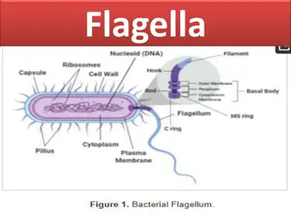
Flagella Slime layer Cytoplasmic membrane Cell wall capsule Nuclear body or nucleoid Cell ribosomes Fimbria mesosome Fig. Structure of a Typical Bacteria
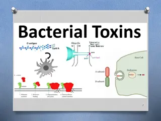
Slime layer and capsule- Extracellular gelatinous accumulation of viscid materials around the external surface of the cell wall of bacteria capsule. The bacterial capsule is a very large structure of many bacteria. It is a polysaccharide layer that lies outside the cell envelope, and is thus deemed part of the outer envelope of a bacterial cell. It is a well-organized layer, not easily washed off, and it can be the cause of various diseases. When it diffuses into the surrounding medium and remains as a loose undermarcated secretion as in leuconostoc, it is known as slime layer.
Function of capsule- 1. Enhances bacterial virulence by inhibiting phagocytosis. Loss of capsule may render the bacterium avirulent. Bacteria tend to lose capsules repeated subcultures. 2. Acts as protective covering against antibacterial substance such as, bacteriophages, phagocytes, enzyme etc. 3. Capsule antigen is specific for bacteria and can be used for identification and typing of bacteria. 4. Diagnostic importance e. g. Mac-Fadyean reaction in Bacillus anthracis and Quelling reaction in case of pneumococcus sp.
Some eg . of Capsulated gram-negative bacteria: Escherichia coli (in some strains) Pseudomonas aeruginosa Salmonella Klebsiella pneumoniae However, some gram-positive bacteria may also have a capsule: Bacillus anthracis Streptococcus pyogenes synthesizes a hyaluronic acid capsule. Streptococcus epidermidis, Staphylococcus aureus
Flagella and Motility-Hair like Helical structure emerge from cell wall.Compsed of flagellin protein and known as H- antigen 1. Monotrichous, e.g. Vibrio cholerae 2.Amphitrichous e.g. Spirillum serpens 3.Lophotrichous e.g. Bartonella bacillifornis 4. Peritrichous e.g. Escherichia coli Fuction- 1. It is the organ of locomotion. 2. Motile aerobic bacteria show positive chemotaxis towards higher oxygen concentration.
3. Motility helps in invasiveness as power of locomotion helps in penetrating through epithelial barrier and viscid mucous membrane. 4.Due to continuous changing of place, the rate of uptake of nutrient solvent is increased. Fimbriae of pili- Some gram-negative bacteria possess another type of filamentous appendages which are numerous, thinner and shorter than flagella.
Functions :------ 1.Ordinarily pili helps in the adherence of symbiotic bacteria to host cell. 2.Pili helps in the attachment of donor and recipient cells in bacterial conjugation and these pili are called sex pili. 3.It helps in the virulence by acting as a colonization antigens. 4. The majority of fimbriae bacteria adhere to red blood cells of many species causing haemagglutination.
Cell wall of bacteria- Rough and rigid structure, 20-25 nm in thickness and weighs about 20-25% of dry weight of the cell. Rigid part is peptidoglycan which is a mucopeptide composed of N- acetyl muramic acid and N-acetyl glucosamine molecules alternating in chains, cross linked by peptide sub units.
Different between gram- negative & gram- pogetive bacterial cell wall
Chemical structure of bacterial cell wall Divided in to four layers- 1. Slime layer and capsule, 2. Surface appendages includes flagella and fimbriae , 3. Cell envelop i.e. Cytoplasmic membrane and cytoplasm containing granular part, nucleoid, ribosome etc. Function of cell wall of bacteria- Shape of the bacteria, Protection to the cell against osmotic damage, Takes part in the cell division, Posses target site for antibiotics, lysozymes and bacteriophages, carries bacterial antigens.
ENDOSPORE An endospore is a dormant, tough, and non-reproductive structure produced by some bacteria in the phylum Firmicutes. The name "endospore" is suggestive of a spore or seed-like form (endo means within), but it is not a true spore (i.e., not an offspring). Bacteria produce a single endospore internally. The spore is sometimes surrounded by a thin covering known as the exosporium, which overlies the spore coat. The spore coat, which acts like a sieve that excludes large toxic molecules like lysozyme, is resistant to many toxic molecules and may also contain enzymes that are involved in germination.
The arrangement of spore layers is as follows: Exosporium Spore coat Spore cortex Core wall The position of the endospore differs among bacterial species and is useful in identification. The main types within the cell are terminal, sub terminal, and centrally placed endospores.
Terminal endospores are seen at the poles of cells, whereas central endospores are more or less in the middle. Subterminal endospores are those between these two extremes, usually seen far enough towards the poles but close enough to the center so as not to be considered either terminal or central. Lateral endospores are seen occasionally. Examples of bacteria having terminal endospores include Clostridium tetani, the pathogen that causes the disease tetanus. Bacteria having a centrally placed endospore include Bacillus cereus. Sometimes the endospore can be so large the cell can be distended around the endospore. This is typical of Clostridium tetani.