Understanding Proteus Species: Morphology, Cultural Characteristics, and Antigenic Structure

Proteus species, specifically P. mirabilis and P. vulgaris, are important opportunistic pathogens in human infections. They exhibit unique characteristics such as swarming growth on agar, gram-negative coccobacilli morphology, and distinctive antigenic structures with O and H antigens. The swarming growth can be inhibited by modifying agar concentration or adding specific chemicals to the media. Understanding the morphology, cultural characteristics, and antigenic structure of Proteus species is crucial for their identification and management in clinical settings.

Download Presentation

Please find below an Image/Link to download the presentation.
The content on the website is provided AS IS for your information and personal use only. It may not be sold, licensed, or shared on other websites without obtaining consent from the author. Download presentation by click this link. If you encounter any issues during the download, it is possible that the publisher has removed the file from their server.
E N D
Presentation Transcript
University of Mustanisiriya College of Medicine Department of Microbiology Third stage Proteus and Pseudomonas spp. Dr Ali Abdulwahid
Proteus spp. Proteus are a member of normal intestinal microbiota and opportunistic pathogens to human The genus belong to the family enterobacteriaceae and divided into four species: P. mirabilis P. vulgaris P. myxofaciens P. penneri P. mirabilis and P. vulgaris are the medically important species as they are causing the majority of infection for human.
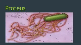
The morphology and cultural Characteristics They are gram-negative coccobacilli They are actively motile with peritrichous flagella and well characterized by swarming growth on agar. Non-motile variants with no flagella can also be present. Non-capsulated They are aerobes and facultative anaerobes. Grow well on ordinary nutrient media . In can be easily recognised by their fishy odor when grow on agar as well as swarming growth on non inhibitory solid media such as nutrient agar and blood agar.
Swarming growth Swarming is an expanding rings or waves of transparent films (composed of bacterial growth) spreading from the margin of a young colony on the surface of the agar plates It is a characteristic feature for P. mirabilis and P. vulgaris and does not happen in the other species Such phenomenon come from the vigorously active motility of the bacterial cells Such growth causes problem in the mixed culture of Proteus with other bacteria, as it may cover most or all of the agar surface as well as colonies of other organisms. Swarming growth of P. mirabilis on agar plate Source of the picture https://commons.wikimedia.org/wiki/Category:Proteus_(bacterium)#/media/ File:ProteusMirabilis_Schw%C3%A4rmen.jpg
Swarming growth The Swarming growth of Proteus can be inhibited by increasing concentration of agar to around 6% or by adding chemicals to the media such as phenylethyl alcohol. The bacteria do not form swarming on MacConkey agar medium, and the colonies are smooth colourless as they NLF (non lactose fermenter) Nonmotile variant do not swarm.
Antigenic Structure Two main antigens O Antigens (Serogrouping) The bacteria were divided into different serogroups depending on their variability in this antigen There are 32 O groups for P. mirabilis and 22 O groups for P. vulgaris In total the two species were divided into 54 O groups on the basis of their O antigens. H Antigens (serotyping) The bacteria were divided intro different serotypes according to the variability in their H antigen There are about 19 different H antigen in P. mirabilis and P. vulgaris were characterised K antigens There is no K antigen as the organism is lack the capsule structure
Pathogenesis Proteus are opportunistic pathogens to humans It commonly causes urinary tract and septic infections, often nosocomial. P. mirabilis are responsible for the majority (90%) of human infection P. vulgaris is a common nosocomial pathogen. Proteus species can cause different infections including : Urinary tract infections (UTI), Wound infections, Pneumonia, Infection of the ear, Respiratory tract infection, Septicemia
Pathogenesis Proteus causes more serious UTI infection than the infection caused by E. coli and other bacteria. The organisms produce the enzyme Urease, which hydrolyse urea into carbon dioxide and ammonia. The ammonia leads to make the urine alkaline, which consequently lead to precipitation of Calcium and Magnesium salts from the urine and producing of urinary stones Ammonia also inactivates the complement and makes damages to the renal epithelium It may also lead to hyperammonaemic encephalopathy and coma. The Proteus bacteria can also colonize the bladder in Patients with long-term indwelling urinary catheters
Laboratory Diagnosis 1. Specimens Specimens are different according to the type of infection and can be include: Urine Blood pus Ear discharge Smear : Gram negative rods can be found in the specimens under the microscope 2. 3. Culture Specimens can plated on different agar media such as MacConkey agar, Nutrient agar, EMB and blood agar The isolate is identified by its morphology and growth features (monitoring the swarming growth or doing gram staining), or doing biochemical and agglutination reactions (using sera targeting O antigens).
Treatment Different strains of Proteus are strongly varying in their susceptibility to antibiotics But they mostly often sensitive to penicillins, cephalosporins, aminoglycosides, and imipenem. The most active antibiotics isaminoglycosides and broad-spectrum cephalosporins P. volgaris in generalmore resistance to some antibiotics (especially ampicillin and amoxicillin)
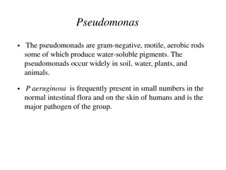
Pseudomonas spp. Pseudomonads group : A large group of Grame ve , aerobic, nons-poring bacilli They are active motile with polar flagella Most are saprophytes found widely in soil, water and other moist environments. Some of them are pathogenic for plants, insects and animals , and few are responsible of infection in humans as opportunistic. They are primarily classified to more than 100 species within the genus Pseudomonas The new taxonomic studies using molecular methods led to allocate many species to new genera such as Burkholderia, Comammona, Stenotrophomonas and Ralstonia
Pseudomonas spp. Pseudomonas aeruginosa is the medically important species that most commonly associated with human disease. This species is widely distributed in nature and is commonly present in moist environments in hospitals. It causes opportunistic infections in different sites in humans with abnormal host defenses.
Morphology and Identification A. Typical organism P. aeruginosa is motile, rod shaped, Gram negative bacilli Can occurs as single bacteria, in pairs, and occasionally in short chains. B. Culture P. aeruginosa grow well on wide range of culture media On nutrient agar, the colonies tend to be large (2-3mm),smooth, translucent, irregularly round. The colonies emit sweet or grapelike odour Some strains produce haemolysin . A gram stain of Pseudomonas aeruginosa. By CDC - Domain, https://commons.wikimedia.org/w/index.php?curid=8099826
Pigments produced by P. aeruginosa There are Four different pigments can be produed by P. aeruginosa include: Pyocyanin : non-fluorescent bluish pigment, diffused into the agar (some species) Pyoverdin : fluorescent pigment gives a green color to the agar ( some strains of P. aeruginosa ) Pyorubin : dark red pigment Pyomelanin: black pigment Produce of glycocalyx capsule Clinical isolates from patients with cystic fibrosis (CF ) often form mucoid colonies on agars due to the overproduction of exopolysaccharide , composed of alginate polymers, known as glycocalyxcapsule Such exopolysaccharide possibly acts as a matrix for the biofilm formation by the organisms Important for evasion of host defenses, especially during chronic pulmonary disease of patients with cystic fibrosis (CF).
P. aeruginosa with fluorescent pigment under UV-light on cetrimid agar-agar. P. aeruginosa with Pyocyanin-pigment on cetrimid agar-agar. Source of the image By BiotechMichael - Own work, Public Domain, https://commons.wikimedia.org/w/index.php?curid=3985161 Source of the image : By BiotechMichael - Own work, Public Domain,https://commons.wikimedia.org/w/index.php?curid=3985120
C. Growth Characteristics P. aeruginosa grows on a wide range of temperature from 6 42 C Grows well at 37 C The ability to grow at 42 C is differential features for P. aeruginosa from other Pseudomonas species in the fluorescent group. They are oxidase positive. Identification is usually based on some feature include: colonial morphology oxidase positivity Pigments production , The ability to grow at 42 C.
Antigenic Structure and virulence factors a) Pili (fimbriae) : mediates attachment to host cells. b) The lipopolysaccharide: variable immunotype, can be used for typing different strains of P. aeruginosa responsible for the endotoxic activity of the bacteria P. aeruginosa can also be typed depending on their susceptibility to the pyocin (bacteriocin). c) Extracellular enzymes: Produced by the majority of the clinical isolates of P. aeruginosa Includes :elastases, proteases, and hemolysins d) Exotoxin A : This toxin lead to necrosis of the tissue and it is lethal for animals when injected in purified form. It is immunogenic, lead to generate antitoxin in human The toxin inhibit the protein synthesis in human cells in the same mechanism of diphtheria toxin
Pathogenesis P. aeruginosa is an opportunistic pathogens They cause infection only when enters cites that lack the normal immune defences in different cases include: Disruption of skin and mucous membrane by direct tissue damage such as in burn wounds. Using of intravenous or urinary catheters. During neutropenia, such as in cancer chemotherapy. The bacterium used their pili to attach to the mucous membranes or skin and colonise them, invades locally, and consequently causes systemic infection . The enzymes and toxins produced by this bacteria promote the local invasiveness and the dissemination of the bacteria . Endotoxic activity of their lipopolysaccharide lead to develop fever, shock, oliguria, leukocytosis and leukopenia.
Pathogenesis The main clinical Findings : 1. wounds and burns infections 2. Meningitis : (route of entry is lumbar puncture or during a neurosurgical procedure) 3. Urinary tract infection: when introduced by catheters and instruments or in irrigating solutions. 4. Necrotizing pneumonia 5. Mild otitis externa in swimmers. 6. Invasive (malignant) otitis externa in patients with diabetes. 7. Eye (Corneal infections) : occurs after injury or surgical procedures, or after using contact lenses and may lead to a rapid destruction to the eye.
Pathogenesis 8. Sepsis: the organisms may invade the bloodstream in patients with leukemia or lymphoma who have received antineoplastic drugs or radiation therapy and in patients with severe burns leading to cause lethal sepsis. 9. Ecthyma gangrenosum : Hemorrhagic necrosis of skin surrounded by erythema and often do not contain pus. It occurs either alone or due to the sepsis caused by P. aerogenosa , especially in people with leukemia and other types of malignant diseases P. aerogenosa can be detected in Gram stained in specimens taken from Ecthyma lesions Culture from such specimens are positive for the organisms The symptoms of infections with P. aeruginosa are varied depending on the infected sites and organs
Diagnostic Laboratory Tests A. Specimens Specimens can be obtained from different body sites depending on the type of infection such as from skin lesions, pus, urine, blood, spinal fluid, sputum etc. B. Smears Microscopic examination of smear can often indicate the presence of Gram-negative rods There are no particular morphological features to differentiate pseudomonas from enteric or other gram- negative rods. C. Culture Blood agar and other differential media used for culturing enteric bacteria can be used for plating specimens of Pseudomonas infections The bacteria can grow readily on these media but tend to be slower than the enteric bacteria The bacteria do not ferment the lactose so it can be easily differentiated from the lactose-fermenting bacteria ( colourless colonies on MacCkonky agar) Culture is the specific test for diagnosis of P. aeruginosa infection ( on nutrient agar, the bacteria form blue-green pigments with fruity odour).
Treatment The bacteria show considerable natural resistance to a wide range of antibiotics When a single antibiotic are used, a rapid resistance can be developed by P. aerogenosa . Therefore combination of an extended spectrum penicillin and an aminoglycoside is recommended. Other clinically effective antibiotics are : carbapenems such as imipenem or meropenem; and the fluoroquinolones, including ciprofloxacin as well as Cephalosporins. The susceptibility patterns of P. aeruginosa vary geographically, and susceptibility tests should be done as an adjunct to selection of antimicrobial therapy.
Epidemiology and Control P. aeruginosa are able to resist to many antibiotics and to grow at wide range of temperature Therefore, the bacteria are able to colonise a variety of environments such as moist equipments in hospital wards, bathrooms and kitchens. The bacteria can also transiently colonise the respiratory and gastrointestinal tracts of hospitalized patients Measures taken to control any other nosocomial pathogens can be also taken to deal with Pseudomonas infections More attention should be paid for sterilizing the hospital equipments, apparatus, tools and dressing Avoiding the excessive and inappropriate use of broad-spectrum antibiotics
References 1. Reidle, S., Morse, S. A., Meitzner, T., and Miller, S. 2019. Jawetz, Melnick & Adelberg s Medical Microbiology , Twenty-Eight Edition. The McGraw-Hill education, Inc. USA 2. Kumar, S. 2012. Textbook of microbiology. Jaypee Brother Medical Publishers (P) Ltd. New Delhi, India.
Thank You For Your Attention