Understanding Tapeworm Infections: Cestodes Overview
Cestodes, commonly known as tapeworms, are long segmented worms with distinct morphology found in the small intestine. This comprehensive guide covers Diphyllobothrium latum, Taenia saginata, their life cycles, morphology, clinical manifestations, diagnosis, and prevention strategies. Detailed information about habitat, epidemiology, infective stages, and key characteristics of these parasites is provided. Understanding the intricacies of tapeworm infections is crucial for effective management and prevention.
Download Presentation

Please find below an Image/Link to download the presentation.
The content on the website is provided AS IS for your information and personal use only. It may not be sold, licensed, or shared on other websites without obtaining consent from the author. Download presentation by click this link. If you encounter any issues during the download, it is possible that the publisher has removed the file from their server.
E N D
Presentation Transcript
CESTODES (TAPEWORMS) All are: 1- Long segmented worms. 2- Same morphology: head (scolex) with or without hooks, have suckers for attachment. short neck. segmented body (proglottid): (immature-mature-gravid segments). 3- Both sexes are in the same body. 4- All found in the small intestine.
DIPHYLLOBOTRIUM LATUM (FISH TAPEWORM) Habitat: small intestine. Epidemiology: World wide - particularly frequent in Scandinavia and northern Canada where raw freshwater fish are consumed. Human is the definitive host. Cyclops are the first intermediate host. Fish is the second intermediate host. Infective stage: plerocercoid.
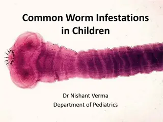
MORPHOLOGY Adult: 10 meters long (3000-4000) segments. Consists of head, immature segments, mature segments and gravid segments. Head: elongated, spoon shape, having two bothria (grooves) Segments: mature and gravid segments have nearly the same morphology. The testes are present in the lateral fields, while the uterus, ovary, commongenital pore are in the center. Egg: oval, operculated, yellowish, immature, 50-70 m.
CLINICAL AND DIAGNOSIS -Diarrhea, abdominal pain -Pernicious anaemia as this worm ingest vitamin B12 leading to its deficiency -Sometimes complications occur in the form of intestinal obstruction. Stool examination for eggs (typical) and segments. Prevention: Freezing at -18oC for 24-48 hrs to kill the plerocercoid Cooking fish. Sewage treatment.
TAENIA SAGINATA (BEEF TAPEWORM) Habitat: small intestine. Epidemiology: Most common tapeworm found world wide. Acquired from eating uncooked beef. Very common in Lebanon and Ethiopia. Human is the definitive host. Cattles are the intermediate host. Infective stage: cysticercous bovis.
MORPHOLOGY Adult: 5 meters long. Consists of head, immature segments, mature segments and gravid segments. Head: has no hooks, has 4 suckers. Segments: Mature segment: The testes are present in the lateral fields, while the bilobed ovary is in the center. Gravid segment: elongated segment, having uterus with lateral branches (more than 14) to accommodate eggs. Egg: mature, spherical, 40 m, brownish in colour, having embryo with six hooks, with thick striated wall.
CLINICAL AND DIAGNOSIS Abdominal pain, diarrhea, loss of body weight, indigestion. Sometimes complication occurs in the form of intestinal obstruction. Stool examination for eggs or segments. Prevention: Good cooking of meat Freeze of meat at -18oC for five days Sewage treatment
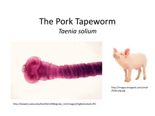
TAENIA SOLIUM (PORK TAPEWORM) Habitat: small intestine. Epidemiology: Not as widely disseminated as T. saginata. Acquired by eating uncooked pork. Human is the definitive host. Pigs are the intermediate host. Infective stage: cysticercous cellulosae.
MORPHOLOGY The same as T.saginata except: Scolex: has 4 suckers and hooks. Gravid segments: number of uterine branches are less than 10 branches.
CLINICAL AND DIAGNOSIS Same as beef tape worm. Prevention: Good cooking of pork meat Freeze of pork meat at -18oC for five days Sewage treatment
TAENIA SOLIUM (CYSTICERCOSIS) Epidemiology: The larval form of T. solium infects humans who ingest the eggs of this tapeworm passed in human stool. Mode of infection: 1-Ingestion of eggs in contaminated food or drink. This occurs most frequently where human stool is used as fertilizer. Also, this occurs most frequently in developing countries where pigs are bred. 2-Autoinfection
Clinical: 1-It is mainly in the skin and the muscles producing mass lesion. 2-The symptoms differed according to site of the cyst, but they are serious when they are present in dangerous area as brain, eyes and heart because of the compressive manifestations. Frequent Presentations are seizures or hydrocephalus. Diagnosis: 1-X-ray to visualize the lesion in the brain and the muscles 2-Serology: IHAT, ELISA 3-Biopsy in case of skin lesion
ECHINOCOCCUS GRANULOSIS: (HYDATID DISEASE) Habitat: Small intestine of dogs. This is a dog tapeworm, the larval (cyst) stage is formed inside the human body which is known as hydatid cyst. Epidemiology: Cosmopolitan. Human is the intermediate host. dogs are the definitive host. Infective stage: eggs.
MORPHOLOGY Adult measures 3-6 mm in length, consists of scolex, neck, one immature segment, one mature segment and one gravid segment.
CLINICAL AND DIAGNOSIS The enlarging cysts can produce symptoms and signs related to their space occupation. It is interfering with the function of the affected organs. Hydatid cysts are dangerous when they occur in the bones, eyes and brain. Rupture of the hydatid cyst results in anaphylactic shock. Also, leakage of scolices and daughter cysts produce hydatid cysts in new locations. 1-Radiology: X-ray, U/S and C.T. are highly effective in locating the cyst. 2-Immunological methods: IHA, CFT, ELISA 3-Eosinophilia: 20-25% 4-Exploratory cyst puncture is contraindicated.
HYMENOLEPIS NANA It is the commonest cestode in children. Man act as intermediate and definitive hosts. Mode of infection: 1-Ingestion of mature eggs in contaminated food or drink 2-Autoinfection. Epidemiology: cosmopolitan. Habitat: small intestine.
MORPHOLOGY Consists of scolex, neck, immature segments, mature segments and gravid segments. Egg: oval or spherical, colourless having two shells, separated by a space. Inside this space, 4-8 filaments. The egg is mature having embryo with six hooks.
CLINICAL AND DIAGNOSIS Abdominal pain, diarrhea, anorexia, headache. stool examination: eggs.

 undefined


