Development of Kidneys and Ureters in Embryology by Dr. Jamila El Medany

This educational material discusses the embryological origin and development of kidneys and ureters. It covers the three systems of kidney development, including the pronephric, mesonephric, and metanephric systems. Detailed descriptions of the formation of the permanent kidney (metanephros), the collecting and excretory parts, as well as common anomalies are provided. The content includes informative images for better understanding.

Download Presentation

Please find below an Image/Link to download the presentation.
The content on the website is provided AS IS for your information and personal use only. It may not be sold, licensed, or shared on other websites without obtaining consent from the author. Download presentation by click this link. If you encounter any issues during the download, it is possible that the publisher has removed the file from their server.
E N D
Presentation Transcript
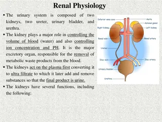
DEVELOPMENT OF KIDNEYS & URETES Dr. Jamila El Medany
OBJECTIVES At the end of the lecture, students should be able to: Identify the embryological origin of kidneys & ureters. Differentiate between the 3 systems of kidneys during development. Describe the development of collecting & excretory parts of permanent kidney. Describe the fetal kidney & identify the pre- and postnatal changes that occur in the kidney. Enumerate the most common anomalies of kidneys & ureters.
EMBRYOLOGICAL ORIGIN from
INTERMEDIATE MESODERM Differentiates into: 1. Nephrogenic ridge (cord) forms kidneys & ureters 2. Gonadal ridge: forms gonads (testes or ovaries)
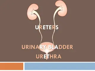
DEVELOPMENT OF KIDNEYS Three systems of kidney develops: 1. Pronephric system: - appears at beginning of 4th week in cervical region - analogous to kidney of fish - formed of tubules & a duct - not function in human - disappears 2. Mesonephric system: - appears at end of 4th week in thoracic & abdominal regions - analogous to kidney of amphibians - formed of tubules & a duct - function temporarily -The duct: In male: forms genital duct - In both sexes: forms ureteric bud 3. Metanephric system: - appears at 5th week in pelvis starts to function at 9thweek Ureteric bud
METANEPHROS (PERMANENT KIDNEY) Formed of 2 origins: Ureteric Bud (derived from mesonephric duct): 1) gives Collecting part of kidney 2) Metanephric Blastema (Mass): derived from nephrogenic cord gives Excretory part of kidney
COLLECTING PART A- Ureteric bud elongates & penetrates metanephric mass. A B- Stalk of ureteric bud forms ureter & its cranial end forms renal pelvis. B C- Branching of renal pelvis gives 3 major calices. Branching of major calyces gives minor calyces. C D- Continuous branching gives straight & arched collecting tubules D
EXCRETORY PART Each arched collecting tubule is surrounded by a cap of metanephric mass. (metanephric vesicle). The metanephric vesicle elongates to form an S- shaped metanephric tubule.
EXCRETORY PART The end of each tubule forms Glomerular (Bowman s) capsule. Each glomerular capsule is invaginated by capillaries (Glomerulus). The tubule lengthens to form: Proximal & Distal convoluted tubules + Loop of Henle
THE NEPHRON (FUNCTIONAL UNIT OF KIDNEY) The Nephron is formed by fusion of: Excretory tubule (from metanephric mass (cap). 1) Arched collecting tubule (from ureteric bud). 2) 2) 1) At Full Term: each kidney contains: 800000 1000000 nephrons.
Criteria of The Fetal Kidney The Kidney is subdivided into Lobes that are visible externally. Lobulation diminishes at the end of fetal period. Nephron formation is complete at birth.
CHANGES of kidney Before Birth Abdominal aorta Common iliac artery Position: The kidney ascends from pelvis to abdomen & attains its adult position, caudal to suprarenal gland. Blood Supply: As the kidney ascends, its blood supply changes from renal branches of common iliac arteries into renal branches of abdominal aorta. Rotation: Initially, the Hilum is ventral then rotates medially about 90 & becomes medial.
What Happens At The 9TH WEEK Beginning of glomerular filtration (start of function). The kidney attains its adult position. Receives its arterial supply from abdominal aorta. The hilum is rotated medially
Changes of kidney After BIRTH 1) Increase in size: due to elongation of tubules and increase in connective tissue between tubules (not due to increase in number of nephrons) 2) Disappearance of kidney lobulation
Congenital Anomalies Pelvic kidney: failure of ascent of one kidney (ureter is short) A. B. Horseshoe kidney: the poles of both kidneys (usually the lower poles) fuse: the kidneys have a lower position than normal but have normal function
B B A- Unilateral renal agenesis: due to absence of one ureteric bud B- Supernumerary kidney: due to development of 2 ureteric buds C- Right side: malrotation of kidney Left side: bifid ureter & supernumerary kidney
GOOD LUCK GOOD LUCK 17