Anatomy of the Kidney: Essential Features and Functions


1
-
Color Index:
Main Text
Male’s Slides
Female’s Slides
Important
Doctor’s Notes
Extra Info
Anatomy of the
Kidney
Renal Block
____________

Objectives
Click
Here
for the
Team’s Summary!
Anatomical features of the kidneys :
position, extent, relations, hilum, peritoneal coverings,
surface anatomy
Internal structure of the kidneys:
Cortex, medulla and renal sinus.
The vascular segments of the kidneys.
The blood supply and lymphatics of the kidneys.
Know the components of the urinary system
Introduction
Functions
1
3
2
6
5
4
Excretes most of the waste products of metabolism.
Controls water & electrolyte balance of the body.
Maintain acid-base balance of the blood.
Erythropoietin hormone stimulates bone marrow for RBCs formation.
Rennin enzyme regulates the blood pressure.
Converts vitamin D to its active form.
1
Only in boys slides
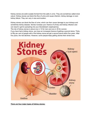
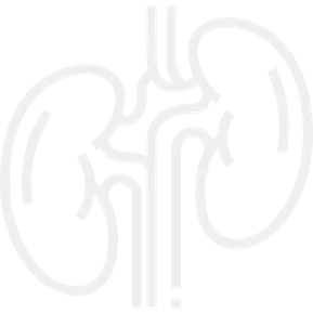
The human body has two kidneys, each around the size of a fist.
They are located at the back of the abdominal cavity just below the rib
cage on each side of the spine.
Every day, each kidney filters liters of fluid from the bloodstream.
Although lungs and skin also play roles in excretion, the kidneys handle the major
responsibility for eliminating nitrogenous (nitrogen-containing) wastes, toxins,
and drugs from the body.
Anatomical Features of the Kidney
2
the kidneys lie in the
retroperitoneal
cavity against the posterior abdominal
wall
on either side of the vertebral
column.
Position
The right kidney lies approximately
1 cm
lower
than the left due to the large size of the right
lobe of the liver.
In the
supine
position, the kidneys extend
from approximately
upper
border of
T12
vertebra
to center of body of
L3
vertebra.
With contraction of the diaphragm during respiration,
both
kidneys move
downward
in a vertical direction, high of one vertebra, 1 inch, 2.5 cm.
Color of the kidney :
reddish brown
Shape:
bean-shaped
Dimensions:
12 x 6 x 3cm.
Although they are similar in size and shape, the
left kidney is
slightly longer and slenderer than the right kidney
, and
nearer to the midline.
Extent
Each kidney has:
●
Convex
upper & lower ends.
●
Convex
lateral border.
●
Convex
medial border at both ends, but
its middle shows a vertical slit called the hilum.
Internally the hilum extends into a large cavity called the
renal sinus.
They
(the kidneys)
are largely under cover of the costal margin.
The upper border of the right kidney is at the level of 12th intercostal
space while the left kidney is at the level of 11th rib.
Hilum and Renal Sinus
3
●
The hilum transmits, from anterior to posterior, the renal
vein
,
renal
artery
& the
ureter
(
VAU
).
and the third branch of renal
artery from the front backward (V.A.U.A.)
●
Lymph vessels & sympathetic fibers also pass through the
hilum.
●
The renal sinus contains the upper expanded part of the ureter called the
renal pelvis.
●
Perinephric fat is continuing into the hilum and the sinus and surrounds
all
these structures.
What Covers the Kidney?
From within outwards the coverings are:
Fibrous Capsule
●
Thin membrane surrounds the kidney and is closely adherent to its
outer surface.
●
Can be easily separated from the surface of the kidney.
Perirenal (Perinephric) Fat
●
Covers the fibrous capsule.
●
The fat is abundant at the borders of the kidney & extends
into the renal sinus.
●
loss of this fat results in nephroptosis kidneys.
Renal Fascia
●
Condensation of areolar C.T that lies outside the
Perirenal
fat and
encloses the kidney and the suprarenal gland.
●
It extends medially and surrounds the large vessels (IVC, aorta), it extends
down and wraps around the ureter and go down towards the pelvis
●
Blood from ruptured kidney or pus from perinephric abscess go down
the renal fascial compartment into pelvis.
●
The midline attachment of renal fascia to the large vessels prevents
inward movement.
Pararenal (Paranephric) Fat
●
Lies external to the renal fascia, is more abundant posteriorly & towards
the lower pole of the kidney.
●
Is part of the retroperitoneal fat.
●
The Perirenal fat, Renal fascia and Pararenal fat
support the kidneys
and hold them in
position
on the posterior abdominal wall.
Relations of the Kidney
Anterior Relations of the kidneys
The anterior surface of both kidneys are related to numerous structures, some with an intervening
layer of peritoneum and others lie directly against the kidney without peritoneum.
Left Kidney
Right Kidney
the anterior surface of left kidney is partly covered by peritoneum:
• A small part of the superior pole, along
the medial border, is covered by
left
suprarenal gland.
• The rest of the superior pole is covered
by the
intraperitoneal stomach and
spleen.
• The
retroperitoneal (body) of pancreas
and splenic vessels
cross the middle part
of the anterior surface.
• Its lower lateral part is directly related to
the
left colic flexure
and beginning of
descending colon.
• Its lower medial part is covered by the
intraperitoneal jejunum.
the anterior surface of right kidney is partly covered by peritoneum:
• A small part of the upper pole is covered
by
right suprarenal gland
.
• The rest of the upper part of anterior
surface is related to the
liver and is
separated by a layer of peritoneum
.
• The
2nd part of duodenum
lies directly
in front of the kidney close to its hilum.
• The lower lateral part is directly related
to the
right colic flexure
and, on its
lower medial side, is related to the
intraperitoneal small intestine.
Posterior Relations of the Kidneys
Posteriorly, the right and left kidneys are almost related to similar structures.
Left Kidney
:
Muscles
:
1- Diaphragm
2- Psoas major
3- Quadratus lumborum
4- transversus abdominis.
Nerves
:
1- Subcostal (T12),
2- iliohypogastric
3- ilioinguinal nerves (L1).
Ribs and recess
:
-
Costodiaphragmatic recess
of the pleura
-
11th
&
12th
ribs; last
intercostal space
4
Right Kidney
:
Muscles
:
1- Diaphragm
2- Psoas major
3- Quadratus lumborum
4- transversus abdominis.
Nerves
:
1- Subcostal (T12),
2- iliohypogastric
3- ilioinguinal nerves (L1).
Ribs and recess
:
-
Costodiaphragmatic recess
of the pleura
-
12th
ribs; last intercostal
space
Vertebrocostal & Renal Angles
The angle between the last rib and the lateral border
of erector spinae muscle is occupied by kidney and is
called the ‘Renal angle’.
The
Vertebrocostal angle
is occupied by the lower
part of the pleural sac.
5
Only in girls slides
Nephron
Each nephron is associated with
two
capillary beds:
●
The glomerulus
●
The peritubular capillary bed.
The glomerulus is both fed and drained by
arterioles
:
-The
afferent
arteriole, which arises from an interlobular artery, is the feeder vessel.
-The
efferent
arteriole receives blood that has passed through the
glomerulus.
Only in boys slides
Internal Structure
Each kidney consists of an outer renal
cortex
and an inner renal
medulla
.
Medulla is
composed of about 12 renal pyramids.
The
renal cortex
is a continuous band of pale tissue that completely surrounds
the renal medulla.
Extensions of the renal cortex, the
renal columns
project into the inner aspect of
the kidney, dividing the renal medulla into discontinuous aggregations of
triangular-shaped tissue, the
renal pyramids
.
Apex, Renal
Papila
The
bases
of the renal pyramids are directed outward, toward the cortex, while the
apex
of each renal pyramid projects inward, toward the
renal sinus.
The apical projection (
renal papilla
) is surrounded by a
minor calyx
.
In the renal sinus, several minor calices unite to form a
major calyx
, and two or
three major calices unite to form the
renal pelvis
, which is the funnel-shaped
superior end of the ureters.
Base
6
Extending from the bases of the renal pyramids into the cortex are
striations known as
medullary rays.
7
Segmental Branches & Vascular
Segments of Kidneys
What is the clinical importance of vascular segments of kidney?
The vascular segments of the kidney provide avascular planes in between
them forming the bloodless line, making it possible to remove individual or
multiple diseased segments or for doing a partial nephrectomy.
Useful in doing incisions for segmental resection of the kidney or opening to
remove a stone from pelvicalyceal system.
Blood Supply of the Kidney
The
renal artery
arises from the
aorta
at the
level of the
second lumbar vertebra.
Together, the renal arteries direct 25% of the
cardiac output towards the kidneys.
The
right renal artery
passes
behind
the IVC.
Abdominal
Aorta
Renal
Artery
Segmental
Arteries
Lobar
Arteries
Interlobar
Arteries
Arcuate
Arteries
Interlobular
Arteries
Afferent
Glomerular
Arterioles
Inferior vena cava
Renal vein
Interlobar veins
Arcuate veins
Interlobular
veins
Arterial Supply
8
Abdominal Aorta
Renal Artery
Segmental Arteries
Lobar Arteries
Interlobar Arteries
Arcuate Arteries
Interlobular Arteries
Afferent Glomerular
Arterioles
Each
renal artery
divides into
5 segmental
arteries that enter the hilum of the kidney,
4 in
front
of the renal pelvis and
one behind
it.
They are distributed to the different segments
of the kidney.
Each segmental artery gives rise to number of
lobar
arteries, each
supplies a renal pyramid
.
Before entering the renal substance, each lobar
artery gives off two or three
interlobar arteries
The interlobar arteries run toward the cortex
on
each side of the renal pyramid.
At the
junction of the cortex and the medulla,
the Interlobar arteries
give off the
arcuate
arteries, which arch over the bases of the pyramids.
The arcuate arteries give off several
interlobular
arteries that ascend in
the cortex and give off the
afferent
glomerular arterioles.
And extend further to become
efferent
arterioles.
Segmental
Arteries
Lobar
Arteries
Interlobar
Arteries
Arcuate
Arteries
Interlobular
Arteries
Arterial Supply Variation
The kidneys present a great variety in arterial supply.
These variations may be explained by the ascending course of the kidney in
the retroperitoneal space, from the original embryological site of formation
(pelvis) to the final destination (lumbar area).
During this course, the kidneys are supplied by consecutive branches of the
iliac vessels and the aorta.
Usually the lower branches become atrophic and vanish while new, higher
ones supply the kidney during its ascent.
Accessory arteries are common (in about 25% of patients).
An accessory artery is any supernumerary artery that reaches the kidney.
If a supernumerary artery does not enter the kidney through
the hilum, it is called aberrant.
Only in boys slides
Lymphatic Drainage and Nerve Supply
Lymphatic Drainage:
-
The lymph vessels follow the arteries.
-
Lymph drains to the
lateral aortic lymph
nodes around
the origin of the renal artery.
Nerve Supply:
-
The nerve supply is the
renal sympathetic plexus
.
-
The
afferent fibers
that travel through the renal plexus
enter the spinal cord in the
10th
,
11th
, and
12th
thoracic
nerves.
Venous Drainage of the
kidney
Inferior vena cava
Renal
vein
Interlobar
veins
Arcuate
veins
Interlobular
veins
9
The Common Developmental Abnormalities of the Kidney:
How to identify that a given kidney is right or left?
-
The hilum is directed medially.
-
The renal vessels are anterior to the pelvis.
-
The ureter is directed inferiorly.
Anterior View
Posterior View
Only in girls slides
Embryology of the Kidney
10
Clinical Notes
It is the preferred terminology for chronic renal failure.
CKD is diagnosed by blood test for creatinine to
determine the patient’s glomerular filtration rate.
High levels of creatinine means the glomerular
filtration rate is falling which means that the kidney’s
ability to filter and excrete waste products is inhibited.
In the early stages of CKD, creatinine levels may be
normal,but urinalysis demonstrates a loss of protein or
red blood cells into the urine.
There are five stages of CKD categorized according to the level of reduced kidney
function and evidence of kidney damage, such as blood or protein in the urine.
The most severe stage is end-stage kidney disease (ESKD), also called end-stage renal
disease and CKD stage 5, which is diagnosed when kidney function deteriorates to the
extent that irreversible kidney failure occurs, requiring kidney dialysis or kidney
transplant.
Only in boys slides
It refers to inability of the kidneys to maintain proper filtration function, excrete wastes
appropriately and to maintain electrolyte balance.
There are three main stages: acute, chronic (now called chronic kidney disease as
discussed above) and end-stage.
Acute renal failure (ARF) is the sudden loss of the ability of the kidneys to remove
waste and concentrate urine.
It is usually initiated by an underlying cause, such as severe dehydration, infection,
trauma to the Kidney or the chronic use of painkillers.
ARF is often reversible with no lasting damage.
ARF is also known as acute kidney injury (AKI).
End-stage renal disease (ESRD) is the complete failure of the kidneys to function, or
where chronic kidney disease has worsened to the point at which kidney function is less
than 10% of normal.
ESRF is also called chronic kidney disease (CKD) stage 5.
See chronic kidney disease above.
11
Clinical Notes
Only in boys slides
It is a kidney disease in which the glomeruli – the parts of
the kidneys responsible for filtering waste and fluids from
the blood - become inflamed.
This causes blood and protein to be lost in the urine.
Glomerulonephritis may be caused by specific problems with
the body’s immune system but often the cause is unknown.
Glomerulonephritis can be acute (a sudden attack of inflammation) - or chronic
(beginning gradually).
In some patients there is no history of kidney disease and the disorder first
manifests as chronic renal failure.
It occurs when the kidneys do not form during fetal
development.
Renal agenesis can be unilateral, with one kidney present,
or bilateral, with no kidneys or very little kidney tissue
present (dysgenesis).
If the agenesis is unilateral, the other kidney will usually hypertrophy to recover for
the missing kidney.
Unilateral agenesis is often asymptomatic and is often discovered later in life.
It involves taking a sample of kidney tissue for laboratory examination.
It can be performed as an open procedure or percutaneously, using a biopsy needle
(generally under ultrasound guidance)
Renal transplantation is a surgical procedure that involves the removal of a diseased
kidney and replacement with a donor organ (either from a living donor or a cadaver).
Living donor kidneys can be either from an identical twin (isograft) or other
individual (allograft), preferably from a close relative.
12
Q1: Mention the FOUR coverings of the kidney?
Answer: Fibrous capsule, perirenal (perinephric) Fat, renal fascia, and pararenal
(paranephric) fat
Q2: List the functions of the kidneys.
Excretes most of the waste products of metabolism.
Controls water & electrolyte balance of the body.
Maintain acid-base balance of the blood.
Erythropoietin hormone stimulates bone marrow for RBCs formation.
Rennin enzyme regulates the blood pressure.
Converts vitamin D to its active form.
Q3: There are 4 muscles related posteriorly to the kidney list them
.
Quadratus lumborum Transversus abdominis Diaphragm Psoas major
Q1: The area where the renal artery enters the kidney
and the renal vein and ureter exits the kidney is called
the
________
.
A- renal hilus
B- cortex
C- medulla
D- renal columns
Q2:
The renal pyramids are separated from each other
by extensions of the renal cortex called ________.
A- renal medulla
B-
minor calyces
C-
medullary cortices
D-
renal columns
Q3: Which of the following structures lies directly in
front of the left kidney?
A-
Quadratus lumborum
B- pancreas
C- Right colic flexure
D-
2nd part of the duodenum
Answers: 1. A 2. D. 3. B 4. B 5. D 6. C
MCQs
SAQs
Q4: Which layer encloses both the kidney and renal
gland?
A- Perirenal fat
B- Renal fascia
C- Pararenal fat
D- Fibrous capsule
Q5: Which of the following is the upper border of the
right kidney?
A- 11th rib
B- 11th intercostal space
C- 10th intercostal space
D- 12th rib
Q6: What artery does glomerular afferent arise from?
A- Arcuate arteries
B- Interlobar arteries
C- Interlobular arteries
D- Lobar arteries
Quiz
Members Board
Team Leaders
Team Members
Mohammad
AlRashed
Najla
AlDhbiban
●
Faisal AlOmar
●
Mishal AlSuwayegh
●
Abdullah AlSalem
●
Saud AlTaleb
●
Abdullah AlNajres
●
Feras AlMasoud
●
Abdulkarim Salman
●
Mayssam AlJaloud
●
Fatima Halawi
●
Ghada BinSlimah
●
Nouf AlDalaqan
●
Shaden AlBassam
●
Maha AlKoryshy
●
Rahaf AlMotairi
●
Haifa AlAmri
●
Alhnouf AlYami
The kidneys play a vital role in filtering waste products, regulating electrolyte balance, maintaining blood pH, producing hormones, and more. This article explores the anatomical features of the kidneys, including their position, extent, internal structure, vascular segments, and coverings. It also delves into the functions of the kidneys in excreting waste, balancing fluids and electrolytes, regulating blood pressure, and activating vitamin D.

Uploaded on Mar 26, 2024 | 2 Views
Download Presentation

Please find below an Image/Link to download the presentation.
The content on the website is provided AS IS for your information and personal use only. It may not be sold, licensed, or shared on other websites without obtaining consent from the author. Download presentation by click this link. If you encounter any issues during the download, it is possible that the publisher has removed the file from their server.
E N D
Presentation Transcript
1 By Faisal Alomar Anatomy of the Kidney ____________ Renal Block Editing File - Color Index: Main Text Male s Slides Female s Slides Important Doctor s Notes Extra Info
Objectives Anatomical features of the kidneys : position, extent, relations, hilum, peritoneal coverings, surface anatomy Internal structure of the kidneys: Cortex, medulla and renal sinus. The vascular segments of the kidneys. The blood supply and lymphatics of the kidneys. Know the components of the urinary system Click Here for the Team s Summary!
1 1 Introduction Only in boys slides The human body has two kidneys, each around the size of a fist. They are located at the back of the abdominal cavity just below the rib cage on each side of the spine. Every day, each kidney filters liters of fluid from the bloodstream. Although lungs and skin also play roles in excretion, the kidneys handle the major responsibility for eliminating nitrogenous (nitrogen-containing) wastes, toxins, and drugs from the body. Functions 1 2 Excretes most of the waste products of metabolism. Controls water & electrolyte balance of the body. 3 4 Maintain acid-base balance of the blood. Erythropoietin hormone stimulates bone marrow for RBCs formation. 5 Rennin enzyme regulates the blood pressure. 6 Converts vitamin D to its active form.
1 2 Anatomical Features of the Kidney the kidneys lie in the retroperitoneal cavity against the posterior abdominal wall on either side of the vertebral column. The right kidney lies approximately 1 cm lower than the left due to the large size of the right lobe of the liver. In the supine position, the kidneys extend from approximately upper border of T12 vertebra to center of body of L3 vertebra. Position Extent With contraction of the diaphragm during respiration, both kidneys move downward in a vertical direction, high of one vertebra, 1 inch, 2.5 cm. Color of the kidney : reddish brown Shape: bean-shaped Dimensions: 12 x 6 x 3cm. Although they are similar in size and shape, the left kidney is slightly longer and slenderer than the right kidney, and nearer to the midline. Each kidney has: Convex upper & lower ends. Convex lateral border. Convex medial border at both ends, but its middle shows a vertical slit called the hilum. Internally the hilum extends into a large cavity called the renal sinus. They (the kidneys) are largely under cover of the costal margin. The upper border of the right kidney is at the level of 12th intercostal space while the left kidney is at the level of 11th rib.
1 3 Hilum and Renal Sinus The hilum transmits, from anterior to posterior, the renal vein, renal artery & the ureter (VAU). and the third branch of renal artery from the front backward (V.A.U.A.) Lymph vessels & sympathetic fibers also pass through the hilum. The renal sinus contains the upper expanded part of the ureter called the renal pelvis. Perinephric fat is continuing into the hilum and the sinus and surrounds all these structures. What Covers the Kidney? From within outwards the coverings are: Fibrous Capsule Thin membrane surrounds the kidney and is closely adherent to its outer surface. Can be easily separated from the surface of the kidney. Perirenal (Perinephric) Fat Covers the fibrous capsule. The fat is abundant at the borders of the kidney & extends into the renal sinus. loss of this fat results in nephroptosis kidneys. Renal Fascia Condensation of areolar C.T that lies outside the Perirenal fat and encloses the kidney and the suprarenal gland. It extends medially and surrounds the large vessels (IVC, aorta), it extends down and wraps around the ureter and go down towards the pelvis Blood from ruptured kidney or pus from perinephric abscess go down the renal fascial compartment into pelvis. The midline attachment of renal fascia to the large vessels prevents inward movement. Pararenal (Paranephric) Fat Lies external to the renal fascia, is more abundant posteriorly & towards the lower pole of the kidney. Is part of the retroperitoneal fat. The Perirenal fat, Renal fascia and Pararenal fat support the kidneys and hold them in position on the posterior abdominal wall.
1 4 Relations of the Kidney Anterior Relations of the kidneys The anterior surface of both kidneys are related to numerous structures, some with an intervening layer of peritoneum and others lie directly against the kidney without peritoneum. Left Kidney Right Kidney the anterior surface of left kidney is partly covered by peritoneum: A small part of the superior pole, along the medial border, is covered by left suprarenal gland. the anterior surface of right kidney is partly covered by peritoneum: A small part of the upper pole is covered by right suprarenal gland. The rest of the upper part of anterior surface is related to the liver and is separated by a layer of peritoneum. The rest of the superior pole is covered by the intraperitoneal stomach and spleen. The 2nd part of duodenum lies directly in front of the kidney close to its hilum. The retroperitoneal (body) of pancreas and splenic vessels cross the middle part of the anterior surface. The lower lateral part is directly related to the right colic flexure and, on its lower medial side, is related to the intraperitoneal small intestine. Its lower lateral part is directly related to the left colic flexure and beginning of descending colon. Its lower medial part is covered by the intraperitoneal jejunum. Posterior Relations of the Kidneys Posteriorly, the right and left kidneys are almost related to similar structures. Right Kidney: Muscles: 1- Diaphragm 2- Psoas major 3- Quadratus lumborum 4- transversus abdominis. Nerves: 1- Subcostal (T12), 2- iliohypogastric 3- ilioinguinal nerves (L1). Ribs and recess: - Costodiaphragmatic recess of the pleura - 12th ribs; last intercostal space Left Kidney: Muscles: 1- Diaphragm 2- Psoas major 3- Quadratus lumborum 4- transversus abdominis. Nerves: 1- Subcostal (T12), 2- iliohypogastric 3- ilioinguinal nerves (L1). Ribs and recess: - Costodiaphragmatic recess of the pleura - 11th & 12th ribs; last intercostal space
1 5 Vertebrocostal & Renal Angles Only in girls slides The angle between the last rib and the lateral border of erector spinae muscle is occupied by kidney and is called the Renal angle . The Vertebrocostal angle is occupied by the lower part of the pleural sac. What care should be taken in exposure of kidneys from behind when 12th rib is to be excised? Push up the pleura which crosses the medial half of the 12th rib. Nephron Only in boys slides Each nephron is associated with two capillary beds: The glomerulus The peritubular capillary bed. The glomerulus is both fed and drained by arterioles: -The afferent arteriole, which arises from an interlobular artery, is the feeder vessel. -The efferent arteriole receives blood that has passed through the glomerulus.
1 6 Internal Structure Each kidney consists of an outer renal cortex and an inner renal medulla. Medulla is composed of about 12 renal pyramids. The renal cortex is a continuous band of pale tissue that completely surrounds the renal medulla. Extensions of the renal cortex, the renal columns project into the inner aspect of the kidney, dividing the renal medulla into discontinuous aggregations of triangular-shaped tissue, the renal pyramids. The bases of the renal pyramids are directed outward, toward the cortex, while the apex of each renal pyramid projects inward, toward the renal sinus. The apical projection (renal papilla) is surrounded by a minor calyx. In the renal sinus, several minor calices unite to form a major calyx, and two or three major calices unite to form the renal pelvis, which is the funnel-shaped superior end of the ureters. Extending from the bases of the renal pyramids into the cortex are striations known as medullary rays. Apex, Renal Papila Base
Segmental Branches & Vascular Segments of Kidneys 1 7 Each kidney has 5 segmental branches and is divided into 5 vascular segments: 1. Apical. (superior) 2. Caudal. (inferior) 3. Anterior Superior. 4. Anterior Inferior. 5. Posterior. What is the clinical importance of vascular segments of kidney? The vascular segments of the kidney provide avascular planes in between them forming the bloodless line, making it possible to remove individual or multiple diseased segments or for doing a partial nephrectomy. Useful in doing incisions for segmental resection of the kidney or opening to remove a stone from pelvicalyceal system. Blood Supply of the Kidney The renal artery arises from the aorta at the level of the second lumbar vertebra. Together, the renal arteries direct 25% of the cardiac output towards the kidneys. The right renal artery passes behind the IVC. Arterial Supply Afferent Glomerular Arterioles Abdominal Aorta Renal Artery Segmental Arteries Lobar Arteries Interlobar Arteries Arcuate Arteries Interlobular Arteries Venous Drainage Interlobular veins Inferior vena cava Renal vein Interlobar veins Arcuate veins
1 8 Arterial Supply Arcuate Arteries Each renal artery divides into 5 segmental arteries that enter the hilum of the kidney, 4 in front of the renal pelvis and one behind it. They are distributed to the different segments of the kidney. Interlobar Arteries Lobar Arteries Abdominal Aorta Renal Artery Each segmental artery gives rise to number of lobar arteries, each supplies a renal pyramid. Segmental Arteries Segmental Arteries Before entering the renal substance, each lobar artery gives off two or three interlobar arteries Lobar Arteries Interlobular Arteries The interlobar arteries run toward the cortex on each side of the renal pyramid. Interlobar Arteries Arcuate Arteries At the junction of the cortex and the medulla, the Interlobar arteries give off the arcuate arteries, which arch over the bases of the pyramids. Interlobular Arteries The arcuate arteries give off several interlobular arteries that ascend in the cortex and give off the afferent glomerular arterioles. Afferent Glomerular Arterioles And extend further to become efferent arterioles. Arterial Supply Variation Only in boys slides The kidneys present a great variety in arterial supply. These variations may be explained by the ascending course of the kidney in the retroperitoneal space, from the original embryological site of formation (pelvis) to the final destination (lumbar area). During this course, the kidneys are supplied by consecutive branches of the iliac vessels and the aorta. Usually the lower branches become atrophic and vanish while new, higher ones supply the kidney during its ascent. Accessory arteries are common (in about 25% of patients). An accessory artery is any supernumerary artery that reaches the kidney. If a supernumerary artery does not enter the kidney through the hilum, it is called aberrant.
1 9 Venous Drainage of the kidney Both renal veins Renal vein emerges from the hilum in front of the renal artery and drain to the inferior vena cava. Kidneys are drained through interlobular veins and interlobar veins until these converge from across the kidney to form the renal vein. The left renal vein - Three times longer than the right (7.5 cm and 2.5 cm) as it courses in front of the aorta to drain into the IVC. - So, for this reason the left kidney is the preferred side for live donor nephrectomy. - It runs from its origin in the renal hilum, posterior to the splenic vein and the body of pancreas, and then across the anterior aspect of the aorta, just below the origin of the superior mesenteric artery. - The left gonadal vein enters it from below and the left suprarenal vein, usually receiving one of the left inferior phrenic veins, enters it above but nearer the midline. (The left renal vein receives left gonadal and left suprarenal veins) - The left renal vein enters the inferior vena cava a little above the right vein. The Right Renal Vein Behind the 2nd part of the duodenum and sometimes behind the lateral part of the head of the pancreas. Venous Drainage Renal vein Interlobar veins Arcuate veins Interlobular veins Inferior vena cava Lymphatic Drainage and Nerve Supply Lymphatic Drainage: - The lymph vessels follow the arteries. - Lymph drains to the lateral aortic lymph nodes around the origin of the renal artery. Nerve Supply: - - The nerve supply is the renal sympathetic plexus. The afferent fibers that travel through the renal plexus enter the spinal cord in the 10th , 11th, and 12th thoracic nerves.
10 1 Embryology of the Kidney Only in girls slides Kidneys develop in the pelvis then migrate up. The Common Developmental Abnormalities of the Kidney: - Aberrant renal arteries: The kidney has more than one renal artery which supply the kidney without passing through hilum. - Pelvic kidney: failure of ascent of kidney from lower lumbar or sacral region. Horseshoe kidney: Fusion of lower poles of two kidneys. It is where the two developing kidneys fuse into a single horseshoe-shaped structure. This occurs if the kidneys become too close together during their ascent and rotation from the pelvis to the abdomen, they become fused at their lower poles (the isthmus) and consequently become stuck underneath the inferior mesenteric artery. This type of kidney is still drained by two ureters (although the pelvices and ureters remain anteriorly due to incomplete rotation) and is usually asymptomatic. - Congenital polycystic kidney. - Congenital absence of one kidney. - Accessory kidney How to identify that a given kidney is right or left? - The hilum is directed medially. - The renal vessels are anterior to the pelvis. - The ureter is directed inferiorly. Kidney location: 1- kidneys extends from T12-L3. 2-kidney hilum is L2 (L1). 3-Ribs 11 & 12 envelop left kidney & Rib 12 envelops right kidney. Anterior View Posterior View
1 11 Clinical Notes Only in boys slides Chronic Kidney Disease It is the preferred terminology for chronic renal failure. CKD is diagnosed by blood test for creatinine to determine the patient s glomerular filtration rate. High levels of creatinine means the glomerular filtration rate is falling which means that the kidney s ability to filter and excrete waste products is inhibited. In the early stages of CKD, creatinine levels may be normal,but urinalysis demonstrates a loss of protein or red blood cells into the urine. There are five stages of CKD categorized according to the level of reduced kidney function and evidence of kidney damage, such as blood or protein in the urine. The most severe stage is end-stage kidney disease (ESKD), also called end-stage renal disease and CKD stage 5, which is diagnosed when kidney function deteriorates to the extent that irreversible kidney failure occurs, requiring kidney dialysis or kidney transplant. Renal Failure It refers to inability of the kidneys to maintain proper filtration function, excrete wastes appropriately and to maintain electrolyte balance. There are three main stages: acute, chronic (now called chronic kidney disease as discussed above) and end-stage. Acute renal failure (ARF) is the sudden loss of the ability of the kidneys to remove waste and concentrate urine. It is usually initiated by an underlying cause, such as severe dehydration, infection, trauma to the Kidney or the chronic use of painkillers. ARF is often reversible with no lasting damage. ARF is also known as acute kidney injury (AKI). End-stage renal disease (ESRD) is the complete failure of the kidneys to function, or where chronic kidney disease has worsened to the point at which kidney function is less than 10% of normal. See chronic kidney disease above. ESRF is also called chronic kidney disease (CKD) stage 5.
12 1 Clinical Notes Only in boys slides Glomerulonephritis It is a kidney disease in which the glomeruli the parts of the kidneys responsible for filtering waste and fluids from the blood - become inflamed. This causes blood and protein to be lost in the urine. Glomerulonephritis may be caused by specific problems with the body s immune system but often the cause is unknown. Glomerulonephritis can be acute (a sudden attack of inflammation) - or chronic (beginning gradually). In some patients there is no history of kidney disease and the disorder first manifests as chronic renal failure. Kidney (Renal) Agenesis It occurs when the kidneys do not form during fetal development. Renal agenesis can be unilateral, with one kidney present, or bilateral, with no kidneys or very little kidney tissue present (dysgenesis). If the agenesis is unilateral, the other kidney will usually hypertrophy to recover for the missing kidney. Unilateral agenesis is often asymptomatic and is often discovered later in life. Renal Biopsy It involves taking a sample of kidney tissue for laboratory examination. It can be performed as an open procedure or percutaneously, using a biopsy needle (generally under ultrasound guidance) Renal Transplantation Renal transplantation is a surgical procedure that involves the removal of a diseased kidney and replacement with a donor organ (either from a living donor or a cadaver). Living donor kidneys can be either from an identical twin (isograft) or other individual (allograft), preferably from a close relative.
Quiz MCQs Q1: The area where the renal artery enters the kidney and the renal vein and ureter exits the kidney is called the ________. Q4: Which layer encloses both the kidney and renal gland? A- Perirenal fat B- Renal fascia C- Pararenal fat D- Fibrous capsule A- renal hilus B- cortex C- medulla D- renal columns Q5: Which of the following is the upper border of the right kidney? Q2: The renal pyramids are separated from each other by extensions of the renal cortex called ________. A- 11th rib B- 11th intercostal space C- 10th intercostal space D- 12th rib A- renal medulla B- minor calyces C- medullary cortices D- renal columns Q6: What artery does glomerular afferent arise from? Q3: Which of the following structures lies directly in front of the left kidney? A- Quadratus lumborum B- pancreas C- Right colic flexure D- 2nd part of the duodenum A- Arcuate arteries B- Interlobar arteries C- Interlobular arteries D- Lobar arteries SAQs Answers: 1. A 2. D. 3. B 4. B 5. D 6. C Q1: Mention the FOUR coverings of the kidney? Answer: Fibrous capsule, perirenal (perinephric) Fat, renal fascia, and pararenal (paranephric) fat Q2: List the functions of the kidneys. Excretes most of the waste products of metabolism. Controls water & electrolyte balance of the body. Maintain acid-base balance of the blood. Erythropoietin hormone stimulates bone marrow for RBCs formation. Rennin enzyme regulates the blood pressure. Converts vitamin D to its active form. Q3: There are 4 muscles related posteriorly to the kidney list them. Quadratus lumborum Transversus abdominis Diaphragm Psoas major
Members Board Team Leaders Mohammad AlRashed Najla AlDhbiban Team Members Faisal AlOmar Mayssam AlJaloud Mishal AlSuwayegh Fatima Halawi Abdullah AlSalem Ghada BinSlimah Saud AlTaleb Nouf AlDalaqan Abdullah AlNajres Shaden AlBassam Feras AlMasoud Maha AlKoryshy Abdulkarim Salman Rahaf AlMotairi Haifa AlAmri Alhnouf AlYami