Types of Placenta in Embryology: Classification and Structure
The lecture discusses the classification of placenta based on the mode of attachment and distribution of chorionic villi. It covers folded, villous, and labyrinthine placenta along with diffuse, cotyledonary, zonary, and discoidal placenta types in different species. The structure of placenta involves six tissues, three from the maternal side and three from the fetal side.

Download Presentation

Please find below an Image/Link to download the presentation.
The content on the website is provided AS IS for your information and personal use only. It may not be sold, licensed, or shared on other websites without obtaining consent from the author. Download presentation by click this link. If you encounter any issues during the download, it is possible that the publisher has removed the file from their server.
E N D
Presentation Transcript
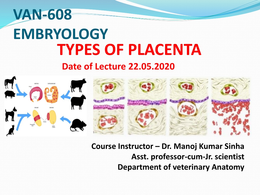
VAN-608 EMBRYOLOGY TYPES OF PLACENTA Date of Lecture 22.05.2020 Course Instructor Dr. Manoj Kumar Sinha Asst. professor-cum-Jr. scientist Department of veterinary Anatomy
TYPES OF PLACENTA The types of placenta is classified on different ways and varied from species to species : 1) Classification on the Basis of Mode of Attachment : It can be classified on the basis of mode of attachment of the chorion to maternal tissue (endometrium) Three types i. Folded Placenta: The surface of chorion and endometrium are interlocked by means of primary and secondary ridges and infoldings Eg. Sow ii. Villous Placenta: The surface of chorion bears villi made up of vascular mesenchymal cones surrounded by trophoblastic and binucleated giant cell. Theses villi either penetrate directly into endometrium Eg. Bitch or simply interdigitate with vascular folds of endometrium eg, Mare and cow , In monkey the chorionic villi are exposed to mternal blood iii. Labryrinthine Plcenta: The chorionic protrusion forming network called chorionic labyrinthine placenta Eg. Canines
continue. 2) Classification Based on Distribution of Chorionic villi : i) Diffuse Placenta :Eg. Horse, Pig, lemur and Camel The chorionic villi are scattered all over the surface of chorion, thus it forms multiple contact between chorion and uterine tissue Cotyledonary placenta : Eg.Ruminants (cud chewing) Cow, Goat,Sheep and Deer . The chorionic villi are present on multiple button like caruncles The tuffts of chorionic villi is called cotyledones which fuses with uterine caruncles to form cotyledonary placenta In ruminants uterus 90-100 carancules are present and arranged into 4 rows which become highly developed on the 30thday of fertilization The uterine caruncles are convex in cow and concave in ewe ii)
Continue.. iii) Zonary Placenta : Eg. Carnivores (Dog and Cat), Raccoon has incomplete zonry placenta The chorionic villi are develop in the form of belt or girdle like band around the middle of their blastocyst or chorionic sac which is elliptical in shape iv) Discoidal Placenta: Eg. Human, Monkey Bat Rodents and Rabbit The chorionic villi are located on a large flattened area in the form of circular disc or plate on the dorsal surface of blastocyst (Monodiscoidal) In Monkey the villi are distributed in two disc, called Bidiscoidal placenta
Continue 3) Structure: Between the maternal and foetal blood there remain a composite layer of tissue In placenta formation six tissue or membranes are participated (3 layer of maternal and 3 layer of foetal tissue ) Three layer of Foetal Side: i) Chorionic epithelium outer layer of foetal membrane derived from trophoblast ii) Connective tissue in the form of chorioallantoic mesoderm iii) Endothelium lining allantoic capillaries Three layer of Maternal Side: i) Endometrial epithelial cells ii) Connective tissue of the endometrium iii) Endothelium lining endometrial blood vessels Classification on the Basis of Histological
Continue.. According to these barriers there are 5 types of Placenta 1. Epithelio-Chorial Placenta : Most primitive type of placenta and all 6 layers are intact Epithelium of uterine mucosa and chorion are lie in apposition to each other At the time of birth the chorion is pulled off the uterine wall without damage of maternal tissue This type of placenta is non-deciduate type Eg. Pig and Mare 2. Syndesmo-chorial Placenta : The uterine epithelium is damaged and stromal tissue of uterus remains in contact with the trophoblast cell of the chorion ,only 5 barriers/tissues are present Eg. Cattle, Sheep, and Deer
Continue 3. Endothelio-Chorial Placenta : The uterine mucosa is reduced and chorionic epithelium comes in contact endothelial the maternal (uterine) blood vessels Only 4 barriers/tissues are presnt between maternal and foetal blood streams Eg. Carnivoers: (dog, cat, bears) with wall of
Continue 4. Haemo-Chorial Placenta: A reduction of three barriers endothelial maternal blood vessels disappear chorionic epithelium is bathed directly maternal blood sinuses Eg. Primates(Humans), Insectivores Shrews), Chiropterans(Bat) i.e. The of walls (uterine) also the and in (Moles,
Continue.. 5. Haemo-Endothelial Placenta: The numbers of barriers between the maternal and foetal blood steram is reduced to 2 The chorionic villi lose their epithelial and connective tissue layer and bare endothelial lining of their blood vessels alone separates the foetal blood from the maternal blood sinuses This is the most intimate type of union and cause extensive hemorrhage during parturition due to loss of both syntrophoblast and cytotrophoblast Eg. Higher Rodents (Rat, Guinea Pig and Rabbit)