Understanding the Anatomy of Ureters, Urinary Bladder, and Urethra
Explore the detailed anatomy of ureters, urinary bladder, and urethra in this comprehensive guide. Learn about the course of ureters, important relations of the urinary bladder, and differences in male and female urethra. Discover the sites of ureteric constrictions, arterial blood supply, and more.

Download Presentation

Please find below an Image/Link to download the presentation.
The content on the website is provided AS IS for your information and personal use only. It may not be sold, licensed, or shared on other websites without obtaining consent from the author. Download presentation by click this link. If you encounter any issues during the download, it is possible that the publisher has removed the file from their server.
E N D
Presentation Transcript
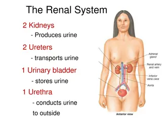
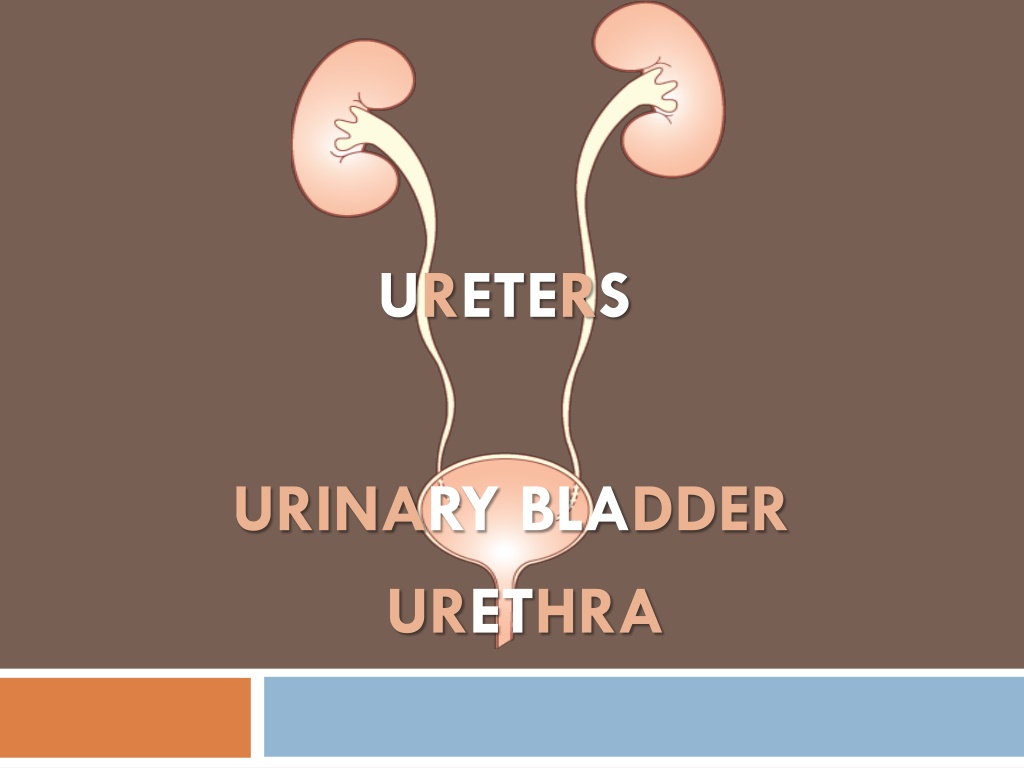
URETERS URINARY BLADDER URETHRA
OBJECTIVES At the end of the lecture, students should be able to: Describe the course of ureter & identify the site of ureteric constrictions. Describe the important relations & identify certain areas (trigone, uvula vesicae) in the base of urinary bladder. List the blood supply, lymphatic drainage & nerve supply of urinary bladder Differentiate between male & female urethra regarding length, structure, course & function.
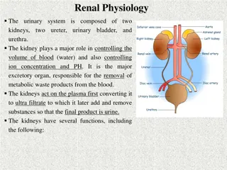
THE URETER Pelvis of Ureter DEFINITION: It is a muscular tube transporting urine from kidney to the posterior surface of urinary bladder. LENGTH: 25 30 cm-(10-12 in) BEGINNING: It begins as a continuation of renal pelvis (or pelvis of ureter).
THE URETER COURSE IN ABDOMEN: It descends anterior to psoas major muscle (opposite the tips of lumbar transverse processes). It crosses anterior to the end (bifurcation) of common iliac artery to enter the pelvis.
THE URETER https://encrypted-tbn2.gstatic.com/images?q=tbn:ANd9GcTCBPggOEmBnCGY81RPf_BJEPybdrd206QkHEtyGW2TCAoLBBSt COURSE IN PELVIS & TERMINATION: It runs downward & backward to the level of ischial spine. It curves forward to open in upper lateral angles of the base of urinary bladder. It runs obliquely for inch in wall of bladder before opening (valve-like part).
THE URETER SITES OF CONSTRICTIONS (OBSTRUCTION-STONE IMPACTION) At ureteropelvic junction At pelvic inlet (site of crossing of common iliac artery) At site of entrance to bladder ARTERIAL SUPPLY: Upper end = Renal artery Middle portion = common iliac artery +testicular or ovarian In pelvis= internal iliac artery
1-THE URINARY BLADDER (SHAPE) It is pelvic organ. It has the shape of three-sided pyramid placed on one of its angle (NECK). It has: An APEX: directed anteriorly (Forward). A BASE: directed posteriorly A SUPERIOR SURFACE Two INFERO-LATERAL SURFACE 1) Picture showing the anatomy of the bladder 2) 3) 4)
2-THE URINARY BLADDER (APEX) Median umbilical ligament Is directed forward. Is related anteriorly to upper border of symphysis pubis. Male Is connected to umbilicus by the median umbilical ligament (remnant of urachus). Median umbilical ligament Female
3-THE URINARY BLADDER (BASE) Lateral veiw Is directed backward IN MALE: Is related to vas deferens & seminal vesicle of both sides IN FEMALE: Male Is related to vagina Posterior veiw Posterior veiw in male Female
4-THE URINARY BLADDER (SUPERIOR SURFACE) IN MALE: Is related to coils of ileum & sigmoid colon Male IN FEMALE: Is related to the uterus Female
5-THE URINARY BLADDER (INFERO-LATERAL SURFACES) Are related to retropubic fat separating them from pubic bones Retropubic fat Accomodates distention of bladder Male Continuous with anterior abdominal wall. Rupture of bladder escape of urine to anterior abdominal wall Female
6-THE URINARY BLADDER (NECK) Is the lowest & most fixed part of urinary bladder. Is continuous with urethra. Is related to (lies behind) lower border of symphysis pubis Male IN MALE: Is related to upper surface of prostate gland (inferiorly, it rests on the base of prostate) Female
7-THE URINARY BLADDER (INTERIOR) Trigone: a triangular area in base of bladder bounded by the 2 ureteric orifices & internal urethral orifice. Its mucous membrane is elastic (not folded) Mucous membrane is folded. Uvula vesicae: elevation behind internal urethral orifice, produced by median lobe of prostate gland
8-THE URINARY BLADDER (CAPACITY) EMPTY DISTENDED Empty bldder is a pelvic organ. Accomodates from 300 500 ml of urine Is circular in shape Bulges into abdominal cavity
9-THE URINARY BLADDER (SUPPLY) ARTERIES: from internal iliac artery http://3.bp.blogspot.com/-LqINgObPWSs/TkoSQl4QKEI/AAAAAAAAACE/qNVYq5G5rho/s400/urinary+bladder.png VEINS: into internal iliac vein Relaxation LYMPH: into internal iliac lymph nodes Cotraction NERVES: Parasympathetic: through pelvic splanchnic nerves from S2, 3, 4 Sympathetic: from L1,2 through hypogastric nerves. Sensory: transmitting pain due to overdistention of bladder (via general visceral afferent fibres from bldder to CNS). 1) Autonomic Regulation of the Bladder 2) 3)
MALE URETHRA (LENGTH: 20 CM) PROSTATIC URETHRA (Length=3 cm): Ejaculatory duct Widest & most dilatable Extends from neck of bladder inside prostate gland MEMBRANOUS URETHRA (Length=1 cm): Surrounded by external urethral sphincter PENILE (SPONGY) URETHRA (Length=16 cm): Structures openings into prostatic urethra: Ejaculatory ducts: containing sperms & secretion of seminal vesicles Ducts of prostate gland Extends inside penis & opens externally through external urethral orifice (narrowest part of whole urethra)
FEMALE URETHRA (LENGTH: 4 CM) Has only urinary function. https://encrypted-tbn2.gstatic.com/images?q=tbn:ANd9GcQ7ie3Ew5pqydqquh87Ad-rmem7XS9dtqtDSIHdWYcgUaUuHI9U Extends from neck of urinary bladder to open externally through the external urethral orifice (anterior to the vaginal opening)
INTRAVENOUS UROGRAM (IVU,IVP) A urogram (Post micturation): demonstrates a bladder stone.Or any obstruction in the urinary system.
SUMMARY-1 URETER: Beginning: as continuation of renal pelvis Course: descends anterior to: psoas major & ends at (bifurcation) of common iliac artery. Termination: opens at upper lateral angle of base of urinary bladder Sites of constriction: at uteropelvic junction, at pelvic inlet, at site of entrance of bladder Arterial supply: renal, gonadal, common & internal iliac arteries
SUMMARY-2 URINARY BLADDER: Apex: related to symphysis pubis, continuous with median umbilical ligament Base: related to vas deferens & seminal vesicle (in male) & to vagina (in female) Superior surface: related to coils of ileum & sigmoid colon (in male) & to uterus (in female) Inferolateral surfaces: related to retropubic fat Neck: continuous with urethra, related to upper surface of prostate gland (in male) Trigone: lies in the base of bladder, bounded by ureteric orifices & internal urethral orifice, its mucous membrane is elastic Uvula vesicae: dilatation behind internal urethral orifice, produced by the median lobe of the prostate gland Supply: internal iliac (artery, vein, lymph nodes) Nerves: parasympathetic (S2,3,4), sympathetic (L1,2) A slight projection into the cavity of the bladder just behind the urethral opening, marking the location of the middle lobe of the prostate gland.
SUMMARY-3 MALE URETHRA: Function: both urinary & genital Length: 20 cm, divided into prostatic (3 cm), membranous (1 cm) & penile ( 16 cm) Course: Extends from neck of bladder to open externally through external urethral orifice (narrowest part of whole urethra) FEMALE URETHRA: Function: urinary only Length: 4 cm Course: Extends from neck of bladder to external urethral orifice (anterior to vaginal opening)