Veterinary Anatomy: Tarsal Bone Structure and Ligaments

Veterinary anatomy lesson featuring the composite joint of the tarsus in an ox, detailing the arrangement of short bones in the tarsus and the ligaments involved. Images and descriptions explain the composition of the composite joint, the types of movements it allows, and the ligaments supporting it, such as the common ligaments, collateral ligaments, and dorsal oblique ligament.

Download Presentation

Please find below an Image/Link to download the presentation.
The content on the website is provided AS IS for your information and personal use only. It may not be sold, licensed, or shared on other websites without obtaining consent from the author. Download presentation by click this link. If you encounter any issues during the download, it is possible that the publisher has removed the file from their server.
E N D
Presentation Transcript
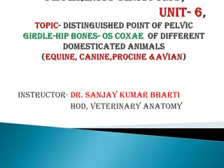
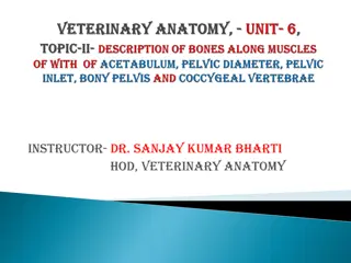
Instructor- DR. SANJAY KUMAR BHARTI HOD, VETERINARY ANATOMY
This is a composite joint consisting of 1. Tibio tarsal articulation 2. Proximal tarsal articulations 3.Inter tarsal articulations 4. Distal tarsal articulations 5.Tarso-metatarsal articulations
TARSAL BONE- OX The tarsus is composed of five short bones arranged in below: Lateral Proximal row Tibial tarsal Medial Fibular tarsal Central row Fused Central and 4th tarsal Distal row First tarsal Fused second & third tarsal
Ox This is a composite joint consisting of 1. Tibio-tarsal articulation 2. Proximal tarsal articulations 3.Inter tarsal articulations 4. Distal tarsal articulations 5.Tarso-metatarsal articulations. The tibio tarsal articulation is a ginglymus or Hinge . The rest are gliding or arthrodia. The joint has common ligaments and special ligaments.
A. Types- Hinge, B-Bone Involvement- Tibia (Distal Extremities) Proximal row)C- Movement- Extension & Flexion D-Common ligaments 1. Capsular ligament may be divided into two parts-anterior and posterior. The anterior capsular ligament is membranous and encloses and tibio- tarsal joint in front. The posterior capsular ligament is also membranous and is attached to the tibial tarsal above, metatarsal below and blends with the lateral ligaments. 2. Collateral Ligaments- The lateral and medial collateral ligaments extending from the distal end of the tibia above on both side lateral and medial, attached to the tarsal bones and the large metatarsal bone below. 3. Posterior ligament (calcaneo-metatarsal ligament) is on the postero- lateral aspect from the posterior border of tuber calcis passes down and is attached to the central and fourth tarsal and to the large metatarsal below. . Tarsal (
4. Dorsal oblique ligament is a narrow band placed at the antero-internal aspect of the joint. It extends from the medial side of the tibial tarsal, passed obliquely downwards and attaches on the fused central, fourth tarsal and the large metatarsal bones 5. Tarso- metatarsal ligament is on the plantar aspect of the joint. It is blended in front with plantar oblique capsular ligament and is adherent to all the tarsal bones. It also blends medially with the medial (collateral) ligament and laterally with planter tarsal ligament. Its plantar face is lines by synovial membrane and forms anterior wall of the tarsal sheath.
ANNULAR certain annular ligament which serve to bind the tendons of the muscles. On the dorsal aspect, there are two-proximal and distal. The proximal annular ligament is large and thick running across from medial to lateral malleolus. It binds fascia down the tendons of complex muscle. The distal annular ligament is thin, arises from fibular tarsal runs obliquely downwards inward is attached to the large metatarsal. The plantar or posterior annular ligament extends from the fibular tarsal and plantar ligament to the medial collateral ligament. This from the posterior wall of the tarsal sheath. The tarsal sheath is formed in front by the tarso-metatarsal ligament (blended with plantar capsular ligament) and behind by the plantar annular ligament. The posterior and anterior faces respectively of these ligaments are lined by synovial membrane and this sheath for the tendon of the deep flexor, plantar arteries and nerves. LIGAMENT 6. -Besides these ligaments, there are
Proximal tarsal articulation- 1.Type- Gliding, 2. Movement- Gliding 3. Bone Involvement-Between tibial tarsal and fibular tarsal bones. 4- Ligaments one extends from the supero-posterior margin of the trochlea of tibial tarsal to fibular tarsal From sustentaculum tali to adjacent part of tibial tarsal on the medial aspect A lateral band from trochlear process of fibular tarsal to lateral ridge of the trochlea of tibial tarsus. An interosseous ligament.
Proximal inter-tarsal articulation 1.Type- Gliding, 2. Movement- Gliding 3. Bone Involvement-Between tibial and fibular tarsals above and central & fourth tarsal. 4.Ligaments Interosseous between tibial and centrals Interosseous between fibular and central tarsals. Distal inter-tarsal articulation1.Type & 2. Movement- Same as above 3. Bone Involvement-Between central & fourth tarsal above fused second and third tarsal and first tarsal below. 4. Ligaments- From central tarsal to fused second and third tarsal on the dorsal aspect. Interosseous ligaments connecting fused central and fourth tarsal to fused second and third tarsal and to large metatarsal.
Distal tarsal articulation- 1.Type & 2. Movement- Same as above 3.Bone involvement- Between fused second and third tarsal and first tarsal 4.Ligament- Interosseous ligament between fused second and third tarsal and first tarsal. Tarsal metatarsal articulation 1.Type & 2. Movement- Same as above 3.Bone involvement-Between central and fourth tarsal, second and third tarsal bones above and large metatarsal bone below. On the dorsal aspect extending obliquely from medial aspect of tibial tarsal, down to large metatarsal. Synovial membranes are four in member. 4.Ligament- Interosseous ligament
Horse Minor differences due to the variation in number of bones. Check ligaments are ill developed. The supra tarsal ligament is vestigial and is represented by deep tarsal fascia, attached to both sides of tendo-achilles. The sub tarsal ligament is the prolongation of the tarso- metatarsal ligament, which joins the deep flexor tendon. Dog & Pig Differences are present due to variation in the number of bones.
FETLOCK JOINT-METATARSO PHALANGEAL ARTICULATION Ox- 1.Type- Hinge, 2. Movement- Extension & Flexion 3. Bone involvement- between the distal extremity of the large metacarpal, the proximal extremity of the first phalanx below and the proximal sesamoids behind. 4. Ligaments The joint has A- capsular, B- collateral and C- sesamoidean ligaments. The capsular ligaments enclose the joints distal extremities of metatarsal over the proximal ends of first phalanges . The collateral ligaments are in the form of axial and abaxial ligaments. The abaxial ligaments extend from the lateral aspects of the distal extremity of the large metatarsal to the abaxial sesamoids and the proximal extremity of the first phalanx below means in a crossing manner . .
The axial ligaments arise common from the cleft of the large metatarsal bone and each spreads out on the cleft of the large metatarsal bone. It then spreads out and attached to the proximal part of the inter digital space of the first phalanx, where its fibres blend with the superior interdigital ligament.
- Sesamoidean ligaments are-4 in number - - A- superior (2 in number) - B- Inter sesamoidean- (3 in number) - C- Collateral (2 in number- lateral & medial ) - D-Inferior-(4 in number in crossing manner two deep and two superficial ) - Total Number of Sesamoidean ligament= 2+3+2+4=11
A- ligament (musculus interossei) arises from the volar face or planter face of the distal row of tarsal bones and the proximal extremity of the large metatarsal descends on the volar face of the latter and divides about its middle into two branches -anterior and posterior.- superior sesamoidean suspensory The or B- Inter sesamoidean- (3 in number) Located between four sesamoid bones like- SB-SL-SB-SL-SB-SL-SB, here total 3 Inter sesamoidean ligamnets present between 4 sesamoid bones
C- Collateral (2 in number- lateral & medial ) Located between distal extremities of Metatarsal and proximal extremity of First Phalanx on its lateral and medial aspect. D-Inferior-or-Distal (4 in number in crossing manner two deep and two superficial ) Located between distal extremities of Metatarsal, tubercle of proximal extremities of first Phalanx


")