Veterinary Guide to Urinary System Examination

This guide covers the examination of the urinary system in veterinary medicine, including collection and analysis of urine samples. It discusses different aspects such as color, volume, transparency, specific gravity, and common interpretations based on findings. Through clear illustrations and detailed descriptions, veterinarians can understand how to interpret urinary system parameters for diagnosis and treatment in animals.

Download Presentation

Please find below an Image/Link to download the presentation.
The content on the website is provided AS IS for your information and personal use only. It may not be sold, licensed, or shared on other websites without obtaining consent from the author. Download presentation by click this link. If you encounter any issues during the download, it is possible that the publisher has removed the file from their server.
E N D
Presentation Transcript
EXAMINATION OF URINARY SYSTEM UNIT -2 B y - D r S o n a m B h a t t A s s i s t a n t P r o f e s s o r V M D B V C P a t n a
COLLECTION OF URINE Free catch (mid stream voided) Catheterisation Cystocentesis Should be analysed as rapidly as possible after collection, ideally within 30 min If this is not possible, samples should be refrigerated immediately & stored preferably for more than 6 12 hours after collection Preservatives: Toluene Thymol Formalin
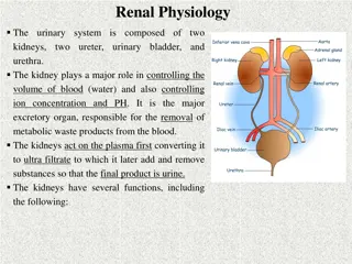
ANALYSIS Physical Physical Examination Examination of Urine Urine: : of Interpretation : Yellow to amber- normal Urine Urine Volume Volume Colorless to pale yellow dilute urine with low specific gravity & polyuria Color Color Recorded while observing it in a test tube or in a urinometer cylinder & & transparency: End stage renal disease Excessive intake of water Diabetes insipidus Normal : yellow to light amber & primarily concentration urochromes Pyometra it depends on the of
Interpretation : Dark yellow to yellow Dark yellow to yellow brown: small quantity brown: concentrated urine with high specific gravity & 1. Acute nephritis 2. Diminished intake of fluids 3. Dehydration prolonged vomiting or diarrhea 4. Fever Red, wine or brown: Hematuria Hemoglobinuria Porphyrins Brown to brownish black: hemoglobin Myoglobin in azoturia Horse: Normal color is yellow when voided, turns deep brown color upon standing (pyrocatechin)
Transparency: Clear, cloudy and flocculent Interpretation Interpretation: : Clear : normal urine, except in horse where it is thick & cloudy due to calcium carbonate crystals & mucus Cloudy : not necessarily pathological 1. Epithelial cells 2. Blood 3. Leucocytes 4. Bacteria 5. Mucus 6. Crystals
Specific gravity: Specific gravity: Directly proportional to urine osmolality, solute concentration and urine density, or the ability of kidney to concentrate or dilute the urine over that of plasma Valuable test, as concentrating ability of the kidneys is among the first sign of renal tubule Recorded either using urinometer method or by a refractometer in cases when the urine quantity is very small NORMAL: 1.015-1.050
Decreased specific gravity Decreased specific gravity Pathologic: Non pathologic: End stage renal disease Excessive water consumption most common cause Diabetes insipidus Diuretics Pyometra Parentral fluids Increased specific gravity Increased specific gravity Pathologic: Dehydration Non pathologic: Fever Decreased water consumption most common cause Diabetes mellitus Disease state in which High environmental temperature abnormal solids are present Excessive panting in urine (protein, glucose)
CHEMICAL EXAMINATION Urinary pH: Urinary pH: Alkaline: horse, cattle, sheep Alkaline: horse, cattle, sheep Acid Acid- - dog, cat dog, cat Acid or alkaline : pig Acid or alkaline : pig Acid- Alkaline- Excess protein Cystitis Starvation Urine retention Prolonged muscular activity Alkalosis Acidosis
Urinary proteins: Tests Tests Robert s test Sulfosalicylic acid test Non pathological causes of proteinuria- high protein meal, exercise and stress Pathological causes include renal disease, glomerular leakage of proteins occur, pyelonephritis, Bence- Jones proteinuria
Glucose: Benedicts test Acetone: Ross test (REAGENT : sodium nitroprusside; ammonium sulfate ) Bilirubin: Gmelin test (nitric acid ): bile duct obstruction, hepatic necrosis caused by infectious canine hepatitis, leptospirosis and other infectious diseases haemolytic diseases such as immune-mediatedhaemolytic anaemia
MICROSCOPIC EXAMINATION Method: Centrifugation Put a sediment on the glass slide Cover with a coverglass Low power microscopy then high power A. Organic sediments: Epithelial cells: squamous epithelial cell; transitional epithelial cells; renal epithelial cells Squamous epithelial cells: largest cell appearing in urine sediment Irregular outline & occur singly or in sheets Derived from urethra & vagina
Significance: Certain number of epithelial cells in urine id normal Squamous epi cells may appear in large number especially in voided samples from female animals Pathologic condition : cystitis, pyelonephritis Erythrocytes : round Leucocyte or pus cells : appear as granular cell that are larger than erythrocyte & smaller than epithelial cells Indications: contamination from genital tract Urethritis Cystitis, pyelitis, nephritis
Upper: RBC in fresh urine are red with a smooth texture. Lower: RBCs in stored urine can crenate taking on a spiky appearance (arrows) WBC; RBC
B. Casts: cylindrical bodies appearing in urinary sediments Formed principally in the lumen of distal tubules & collecting tubules of kidney When cells & cellular debris are present in the tubules, they are included in hyaline matrix at time of formation Types of casts: Hyaline cast: made of protein alone Colorless, homogenous, semitransparent, cylindrical structures with parallel sides & usually rounded ends Indicates mildest form of renal irritation
Granular cast: hyaline cast containing fine or coarse granules ; chiefly derived from disintegration of tubular epithelial cells Most common type of cast seen in domestic animals Indicates a more severe type of renal disease than hyaline cast, represent integration of renal tubular epithelium Large numbers are usually associated with more severe renal disease involving necrosis of tubular cells Epithelial cast:, waxy cast, fatty, blood, leucocytic, mucus & mucus thread Significance: Generally indicates degree of renal changes renal irritation. Renal inflammation, renal degeneration Microorganisms: bacteria, yeast, fungi, parasite
Unorganised sediment: 1. Crystals: Depends on pH Normal: Acid urine: amorphous urates & uric acid Alkaline urine: triple & amorphous phophates, calcium carbonate Abnormal: minor significance; except in case of urolithiasis & few metabolic disturbance
Struvite (coffin shaped) Calcium carbonate
Hyaline cast Granular cast


")