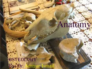
Veterinary Anatomy: Pelvic Structures and Sexual Dimorphism in Animals

The article discusses the anatomy of the acetabulum, obturator foramen, pelvic cavity, pelvic inlet and outlet, and sexual differences in the pelvis, with a focus on sheep and goats. It covers the structure of these pelvic components and highlights the variations between male and female anatomies. Detailed descriptions and images accompany the informative content.

Download Presentation

Please find below an Image/Link to download the presentation.
The content on the website is provided AS IS for your information and personal use only. It may not be sold, licensed, or shared on other websites without obtaining consent from the author. Download presentation by click this link. If you encounter any issues during the download, it is possible that the publisher has removed the file from their server.
E N D
Presentation Transcript
Instructor- DR. SANJAY KUMAR BHARTI HOD, VETERINARY ANATOMY
Acetabulum Acetabulum is a cotyloid cavity formed on the ventro-lateral aspect of the os coxae by the meeting of its three components. It consists of an articular and a non-articular part. The former is nearly circular and articulates with the head of the femur. The rim of the cavity presents on its postero-medial spect the acetabular notch, which transmits the round ligament of the hip joint. The non-articular part, the acetabular fossa is situated in the depth of the acetabulum. Another small notch may be seen antero-medially, though sometimes it is replaced by a foramen or is absent.
Obturator foramen The obturator foramen is a largest foramen of body, elliptical opening on the floor of the pelvis and is circumscribed by the ischium and the pubis. It is covered in life by the obturator muscles. Pelvic cavity The pelvic cavity is the smallest and the most posterior of the three visceral cavities of the body. The bony pelvis composed of the os coxae laterally and ventrally. The three coccygeal vertebrae dorsally. The lateral vacuities are closed up by the sciatic ligaments in life. sacrum and the first
The pelvic inlet is bounded by the terminal line or brim which is composed of the base of the sacrum dorsally, laterally and the anterior borders of pubis ventrally. The pelvic outlet is much smaller and is very incomplete in the skeleton. The third coccygeal vertebra bound it dorsally and ventrally by the ischial arch and the sacro-sciatic ligament and semimembranosus muscle completes it laterally. ilio-pectineal lines
Sexual Sexual differences differences The ischial arch is wider and the outlet is larger in the female than in the male. The conjugate (vertical) and transverse diameters are greater in the female so that the cavity is roomier. The pubis and the ischium of the opposite sides meet at a more open angle in the female than in the male.
Sheep and Goat 1.The long axis of the ilium is almost in straight line with that of ischium. 2. Greater ischiatic spine is low and everted. 3. Ischial symphysis is not ossified even in old animals. 4. Acetabulum is large and deep. 5. The pelvic brim is elliptical in outline.
1. The gluteal line is very faint or not prominent . 2. This bone inclined forward in position but tuber sacral inclined backward. 3.The Superior ischiatic spine is not sharp. 4.The Psos tubercle is under developed. 5. The cotyloid cavity is wide &Acetabular notch is also wide & medial in position. 6. Supra acetabular fossa is not prominent. 7. The ischial arch is wide and shallow. 8. The ridge on the inferior face of the ischium is absent. 9. The symphyseal ridge is also absent. 10. The tuber ischii is not trifid. 11. The ventral face of the pubis transmits the pubo-femoral or the accessory and round ligaments of the hip to femur. 12. The Ventral tubercle is absent.
1.The ilium is parallel with opposite.. 2. The ilium and ischium are almost in line with each other . 3. The gluteal surface is concave. 4.The Crest of ilium is more convex. 5.Superior Ischiatic spine is blunt. 6.The ilio-pectineal line is prominent and the psoas tubercle is well marked 7.The Ischium is twisted. 8.The tuber ischii or Ischial tuberocity is flat. 9. The Ischial arch bifurcated. 10. The obturator foramen somewhat complete Oval in shape.
1. Os coxae is long and narrow. 2. The ilium and ischium are almost in line with each other. 3. The gluteal surface is divided into two fossa by a ridge which is continuous with the greater ischiatic spine behind. 4. The iliac crest forms the highest point of the bone. 5. There is a crest or tubercle on the ventral surface of the ischium. 6.Acetabular notch is thicker. 7.The ilio-pectineal line is prominent and the psoas tubercle is well marked. 8. Pelvic inlet is elliptical in outline.
The ilium is elongated and extends over the entire length of the hipbone. It is firmly fused to the transverse processes of the lumbo- sacral mass. Means ilium bone fused with sacrum. The ischium is smaller and lies below and lateral to the posterior part of the ilium is triangular. The sciatic foramenor ilio-ischiatic is formed by the adjacent borders of the ischium and ilium behind the acetabulum . The ventral border forms the obturator foramen with the pubis. The pubis is a long and slender rod running along the ventral border of the ischium. The anterior end has a muscular process. The acetabulum is large and perforated. 1. 2.
BONE Bony pelvis consist of sacrum, 1stthree coccygeal vertebrae and two Os- coxae. Each formed by the ilium, ischium and pubis. Sacrum S. No. Mare Cow 1. It consist of 5 sacral segments, 1. It consist of 5 sacral segments. base and apex. 2. It is shorter than the cow 2.It is longer than the mare. 3. Triangular in shape. 3. Roughly triangular in shape. 4. Dorsal surface presents 5 sacral spines 4. Dorsal surface presents spines fuse which have tuberous summits and to form median sacral crest 4 pairs of dorsal sacral foramina. Articular processes fuse to form lateral sacral crest and 4 pairs of dorsal sacral foramina.
BONE SACRUM S.N. MARE COW 5. Ventral surface lodges 4 transverse lines. 5. Ventral surface presents 4 transverse groove and 4 pairs of foramina 6. Pelvic surface presents transverse line 6. Concave in both direction having groove for medial sacral artery 7. Smooth notch are present 7. Absent 8. Articular surface directed 8. Articular surfaces are concave dorso-laterally 9. It is rough, irregular and articulate 9. emicylindrical in curvature medially with ilium.
Sacro-pubic (Conjugate) diameter :- It is measured from sacral promontory to the anterior margin of pubis symphysis. Transverse (Bis-iliac) diameter :- Measured at its greatest width just above psoas tubercle. Superior bis-iliac diameter :- It is measured at upper 3rdof pelvic inlet and receives greatest width of the fetus at shoulder in anterior presentation and hip in the posterior presentation. Inferior bis-iliac :- It is measured at the lower 4thof pelvic inlet at elbow of fetus in anterior presentation and stifle in posterior presentation. Vertical diameter of inlet :- It is measured between anterior end of symphysis pubis and articulation of sacral 3rdand 4thvertebra. Oblique / sacro-iliac / ilio-sacral diameter of inlet :- It is measured from sacro- iliac joint of one side through the center of pelvic cavity to the psoas tubercle of opposite side. It is intermediate between sacro-pubic and superior bis-iliac diameter
Coccygeal vertebrae Each vertebra consist of body, spinous and transverse processes. S.No. Cow 1. 18-20 in numbers 1. 15-21 in number 2. 1stVertebrae, well developed 2 . Having groove medially 3. Ventrally groove for median 3. For coccygeal artery saccral artery. 4. First 5-6 vertebare having 4. Less Developed complete arches and spinous process. Mare


 diameter :- It is measured from")