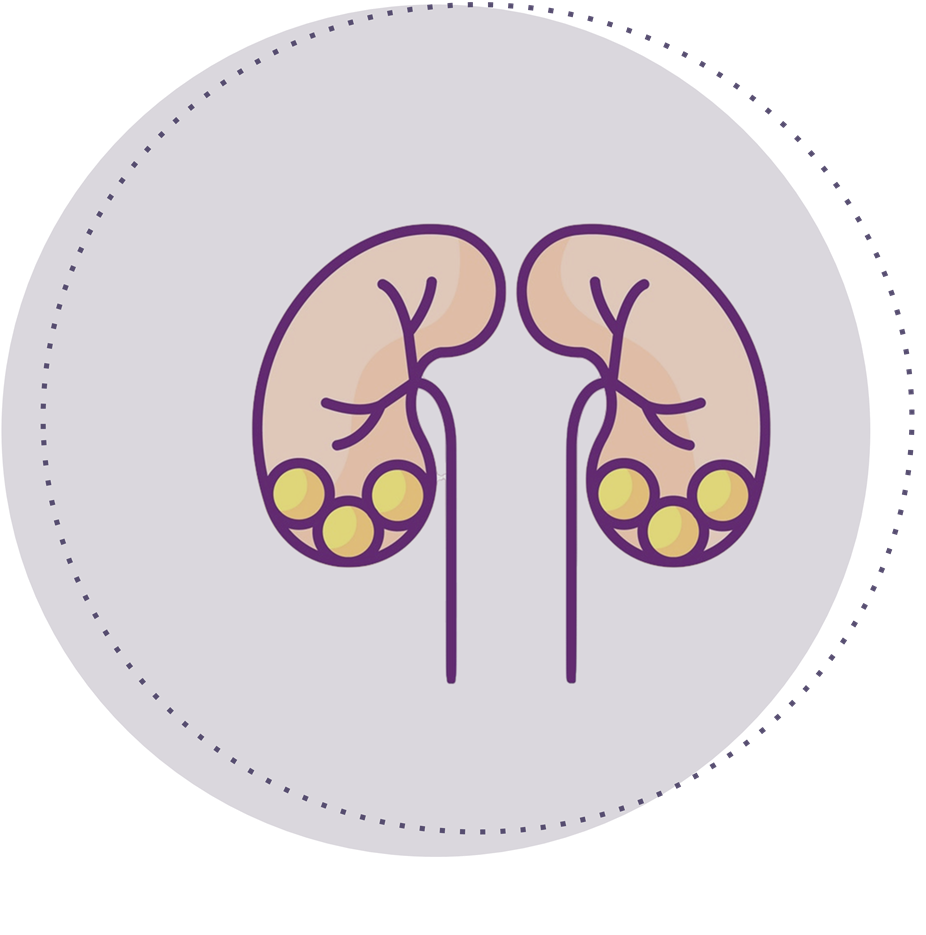
Pathology Acute kidney injury


Pathology

Acute kidney injury


Editing File

- Color Index:
Main Text
Male’s Slides
Female’s Slides
Important
Doctor’s Notes
Extra Info


L1


Upon completion of this lecture the students will be able to:
➔
Describe the guidelines of the renal biopsy.
➔
Recognize the types of acute kidney injury.
➔
Recognize the clinical manifestations of acute kidney injury.
➔
Describe the pathological findings in acute kidney injury.
Key Outlines:
➔
Brief review of the normal anatomy and histology of the kidney
and urinary tract.
➔
Terminology.
➔
Etiology.
➔
Pathophysiology.
➔
Clinical manifestations with diagnostic approach.
➔
Pathological evaluation: The four elements possibly implicated:
Gross and histological findings.
➔
Conclusion.


Objectives


Introductory slide
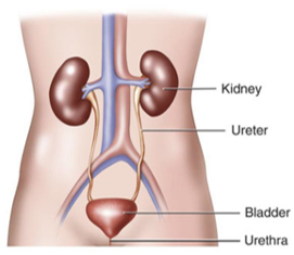


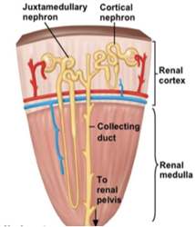
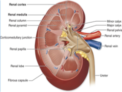
Anatomy of the kidney
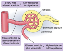
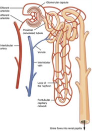


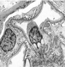
Histology of the glomerulus


Under the
(EM)

Under the
(LM)


Under the
(EM)

Under the
(LM)

Scanning EM

Transmission
(Cross section)
EM

Excretory function:
Detoxify blood, removal of toxins, nitrogenous wastes (urea
and creatinine), drugs etc.
Homeostatic function:
●
Maintain and regulate water balance and osmolality.
●
Maintain and regulate electrolyte balance (Na+, K+,
Ca2+, Cl-, Mg2+ ions etc) .
●
Maintain and regulate acid-base balance (pH, H+ and
HCO3- ions).
Endocrine function:
●
Secretes
renin
from the JGA cells to regulate BP and
electrolyte balance.
●
Secretes
erythropoietin
from the endothelial cells lining the
cortical peritubular capillaries to stimulate the bone
marrow to produce RBCs.
●
Converts inactive Vit D3 to active 1,25
dihydroxycholecalciferol by alpha one hydroxylase enzyme
under the influence of PTH (Parathyroid hormone) →
increases calcium absorption.
Normal Kidney Function
Uremia
Azotemia
Oliguria
Anuria
●
is a clinical syndrome associated with fluid,
electrolyte, and hormone imbalances and
metabolic abnormalities, which develops
with deterioration of renal function.
●
due to the accumulation of organic waste
products that are normally cleared by the
kidneys.
●
uremia means urine in the blood.
Uremia can be seen in both chronic kidney
disease and acute kidney injury.
●
Is abnormally high levels of nitrogen-
containing compounds
(such as urea,
creatinine etc) in the blood.
●
It can lead to uremia if not controlled.
●
It is an
elevation of blood urea nitrogen
(BUN) and serum creatinine levels.
●
Urine output is
markedly
less than 400
ml/24 hours
●
Urine output
less than 200 ml/24
hours
Important terminology
Renal Failure
(
Insufficiency)
●
is when the kidneys fail to adequately
filter toxins and waste products from the
blood.
●
In renal failure there is
decrease
in
glomerular filtration rate and an
elevated
serum creatinine level.
Note
: Creatinine clearance or filtration is
dependent on the glomerular filtration rate (GFR).
The tissue obtained is used to make slides for:
Light microscopy (LM) :
to study the histology in renal cortex & medulla.
study is to detect:
the presence of immunoglobulins
(IgA, IgG, IgM)
and complements (
C3
and
C1q
) in the glomerular mesangium or in the wall of the glomerular
capillary loops.
Immunofluorescence (IF) :
Electron microscopy (EM)
(ultrastructural) :
study is to detect the presence or absence of:
●
effacement of the epithelial cell (podocytes)foot
processes.
●
electron dense immune complex deposits
●
If deposits are present then to identify the location of the
deposits in the glomeruli (mesangial/paramesangial,
subepithelial, subendothelial).
Kidney biopsy
Renal failure classification
Based on Duration
Chronic
Acute
●
Sudden onset.
●
Rapid reduction in urine
output.
●
Usually reversible.
●
Gradually progressive
with nephron loss.
●
Usually not reversible.
Based on Etiology
1- Pre-Renal
2- Renal
3- Post-Renal
(55-60%)
The renal tubular
and glomerular
function are normal.
Here the GFR is
decreased due to
reduced renal
perfusion.
(35-40%)
Is due to diseases
of the kidney itself
(which is
associated with
release of renal
afferent
vasoconstrictors).
(5-10%)
Occurs when
obstruction in the
urinary tract below
the kidneys causes
waste to build up
in the kidneys.
Based in the urine output
Oliguric
Non-oliguric
Anuric
Urine output less
than 400ml/day
Urine output
greater than
400ml/day
Urine output less
than 100ml/day
NOTE:
sometimes more than 70% of renal function
can be lost before it is clinically noticeable.
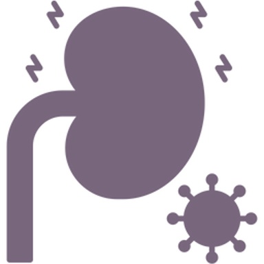
Acute kidney injury
Definition
AKI is a syndrome defined by a
sudden loss of renal
function over several hours to days
resulting in the
accumulation of nitrogenous compounds such as urea and
creatinine.
It is characterized by:
Sudden
decrease
in
GFR (hours
to days)
Accumulation of
nitrogenous waste
products.
Rapid rise
in serum
creatinine
Oliguria: the
urine output
is markedly
decreased
(usually it is
less than 400
ml/day)
Fluid
imbalance;
electrolyte
imbalance;
acid-base
disturbance;
and mineral
disorders.
(previously called acute renal failure [ARF])
Note
: the
contra
st
mediu
m/dye used in
variou
s radiological tests can
occasi
onally induce
AKI.
Note:
In AKI if there is reduction in renal blood flow/ renal perfusion
decrease in glomerular filtration rate (this is the common pathologic
pathway for AKI regardless of the cause). The decreased GFR
activation of renin-angiotensin-aldosterone system. These physiologic
changes result in increased sodium reabsorption, increased water retention,
increased urinary creatinine concentration, increased urine specific gravity
and increased urine osmolality. Acids are retained and HCO 3 is excreted.
Pathogenesis Of Acute Kidney Injury
1- Pre-Renal azotemia
Is the
most
common
cause of
acute
renal
failure
(ARF).
It results from
Decreased renal
perfusion/hypope
rfusion
(i.e.
decreased
effective blood
flow to the kidney.
Initially the body
tries to
compensate but
in
severe
hypoperfusion
→
GFR is markedly
decreased → AKI
The treatment
of the cause
Restores
the
renal function.
Pre Renal causes of ARF are:
Anything
that leads to renal
Hypoperfusion
:
Impaired cardiac function (cardiac failure, myocardial infarction, massive
pulmonary embolism).
Cirrhosis (it can cause renal vasoconstriction → kidney injury called
“hepatorenal syndrome”)
Anaphylaxis.
Hypovolemia
: e.g. hemorrhage, volume depletion (dehydration or GIT fluid
loss in vomiting, diarrhea), hypoalbuminemia, diuretics, third space losses
(burns, peritonitis, muscle trauma) etc.
Sepsis, septic shock.
Other causes: surgery, NSAIDS and other nephrotoxic drugs etc.
Based on Kidney Disease Improving Global Outcomes(KDIGO) AKI is
defined by the presence of any one of the following:
(important to know all of them because it may come as cases) + MCQs
3- Post renal causes of AKI/ ARF
Any obstruction to the outflow of urine:
Congenital or structural abnormality
Benign prostatic hyperplasia (older men)
Stones in the urinary tract
A tumor in the urinary tract (e.g. ureter, bladder, prostate, urethra).
External compression from the outside of the urinary tract (e.g.
retroperitoneal fibrosis or tumors like carcinoma of cervix).
2- Renal causes of AKI (intrinsic)
The kidney biopsy of a patient with AKI can show any of the previously
mentioned causes.
The common causes are:
Note: Common causes of AKI
Acute tubular injury and Acute tubular necrosis
Tubular cause:
acute tubular injury/ acute tubular necrosis
Glomerular cause:
Interstitial cause:
acute tubulointerstitial nephritis
(later lectures)
Post infectious glomerulonephritis
(later
lectures)
Rapid progressive crescentic
glomerulonephritis
(later lectures)
●
bilaterally enlarged & swollen kidneys
(due to edema)
●
Cut surface shows a pale cortex and a
dark & congested medulla
Gross morphology
●
Ranges from tubular epithelial cell
swelling
(mild injury) to tubular epithelial cell
necrosis
(severe injury).
●
The proximal tubules become
dilated
and
irregular in shape
●
The tubular epithelial cells become
flattened
(so the
wall of the tubule becomes thin
)
and show
loss of brush border.
●
In the necrotic tubular epithelial cells there is
loss of nucleus
and also there is sloughing
(falling off) of necrotic cells into the lumen of
the tubule.
●
Casts may be present in the lumen of the
distal tubules.
●
Interstitium
→
mild edema.
●
Later as healing begins there is regeneration
of the tubular epithelial cells
Histology
●
Oliguria
: Decreased urine output (occasionally urine output remains
normal). Typically the urinary output is < 400 ml/day.
●
Defective elimination of metabolic waste, water, electrolytes, and acids
from the body. Waste material is accumulated in the body which causes
azotemia
(> nausea & vomiting)
, acid-base imbalance
(> acidosis)
and
electrolyte imbalance like
hypernatremia and hyperkaliemia (>
abnormal heart rhythms, risk of heart failure, weakness and
muscle paralysis).
●
Salt and water retention >
generalized
edema
starting with swelling in
your legs, ankles or feet.
●
Pulmonary edema
(shortness of breath due to extra fluid on the lungs).
●
Other features: hypotension, nephritic syndrome, flank pain, fatigue,
uremic encephalopathy, confusion, seizures or coma in severe cases
●
Sometimes acute kidney failure causes no signs or symptoms and is
detected through lab tests done for another reason.
Clinical features of AKI
Treatment
Treat the underlying etiology.
Dialysis
Correction of fluid imbalance.
Correction of acidosis and electrolyte
imbalance e.g hyperkalemia
Summary of causes of AKI :
-Bilaterally enlarged & swollen kidneys (due to edema).
-Cut surface shows a pale cortex and a dark & congested medulla.
• Ranges from tubular epithelial cell
swelling
(mild injury) to tubular epithelial cell
necrosis
(severe injury). • The
proximal tubules are dilated
with
thinning of tubular wall
. • The
tubular epithelial cells are
flattened
and show
loss of brush border
• Hyaline, granular and
pigmented casts are present in the lumen of the distal tubules. • The interstitium has
mild
edema
•Later as healing begins there is subsequent regeneration of the tubular epithelial cells
prolonged
ischemia to
nephrons
leads to
tubular
injury and
necrosis.
Endogenous toxin
Pigments and
casts:
- Myoglobinuria
(in
Rhabdomyolysis)
- Hemoglobinuria
- Myeloma casts
etc.
Exogenous toxin
Nephrotoxic drugs:
- Aminoglycosides
(antibiotic).
- Amphotericin B
- Calcineurin inhibitors
(e.g.,tacrolimus) etc.
Radiographic contrast
medium
(dyes induced toxicity)
Toxicity to nephrons
leads to tubular injury
& necrosis
Toxicity to
nephrons leads to
tubular injury &
necrosis
A.
Pre-Renal cause
B.
Post-Renal cause
C.
Renal cause
D.
Intrinsic cause
Q
1
)
M
o
s
t
c
o
m
m
o
n
e
t
i
o
l
o
g
y
o
f
r
e
n
a
l
f
a
i
l
u
r
e
:
A.
Oliguric
B.
Non-oliguric
C.
Anuric
D.
None of the above
A.
Uraemia
B.
Azotemia
C.
Anuric
D.
Oliguria
A.
LM
B.
EM
C.
IF
D.
Ultrasound
Quiz
Q
2
)
W
h
e
n
t
h
e
u
r
i
n
e
o
u
t
p
u
t
i
s
l
e
s
s
t
h
a
n
4
0
0
c
c
/
2
4
h
r
i
t
’
s
:
Q
3
)
a
b
n
o
r
m
a
l
l
y
h
i
g
h
l
e
v
e
l
s
o
f
n
i
t
r
o
g
e
n
-
c
o
n
t
a
i
n
i
n
g
c
o
m
p
o
u
n
d
s
Q
4
)
I
s
s
t
u
d
y
i
n
g
t
h
e
h
i
s
t
o
l
o
g
y
i
n
r
e
n
a
l
c
o
r
t
e
x
&
m
e
d
u
l
l
a
.
Q
5
)
T
r
e
a
t
m
e
n
t
o
f
a
c
u
t
e
t
u
b
u
l
a
r
i
n
j
u
r
y
a
n
d
a
c
u
t
e
t
u
b
u
l
a
r
n
e
c
r
o
s
i
s
i
n
c
l
u
d
e
s
a
l
l
b
u
t
t
h
e
f
o
l
l
o
w
i
n
g
.
Q
6
)
I
n
t
y
p
i
c
a
l
p
a
t
i
e
n
t
w
i
t
h
a
c
u
t
e
k
i
d
n
e
y
i
n
j
u
r
y
w
h
i
c
h
f
e
a
t
u
r
e
i
s
m
o
s
t
l
i
k
e
l
y
t
o
b
e
p
r
e
s
e
n
t
.
A.
Treat the underlying etiology
B.
Dialysis
C.
Correction of fluid imbalance.
D.
surgery
A.
Oliguria ( urinary output is < 400 ml/day)
B.
fever
C.
coma
D.
hypokalemia
Answer: Q1/A Q2/A Q3/B Q4/A Q5/D Q6/A Q7/ C
Q
7
)
W
h
i
c
h
o
n
e
o
f
t
h
e
f
o
l
l
o
w
i
n
g
c
a
u
s
e
n
e
p
h
r
o
t
o
x
i
c
i
n
j
u
r
y
?
A.
hemorrhage
B.
incompatible blood transfusions
C.
Radiographic contrast agents
D.
dehydration
●
Hoor Aloraini
●
Farah alhalafi
●
Shahad Alsalahi
●
Nouf Aldhalaan
●
Jana alhazmi
●
Mayssar Alshobaki
●
Rahaf Almutairi
●
layan Aldoukhi
●
Atheer Alahmari
●
Walaa AlMutawa
●
Fatima Halawi
Members
Members
Team Leaders
Hassan Alabdullatif
Team Members
Theme was done by Shatha Almutib
Maha Alkoryshy
Manar Alzahrani
●
Omar Alkadhi
●
Emad Almutairi
●
Rakan Alromayan
●
Ali Alzahrani
●
Saad Alahmari
●
Malik halees
●
Khalid Alrasheed
●
Nawaf Alturki
Pathology442@gmail.com
Guidelines of renal biopsy, types of acute kidney injury, clinical manifestations, and pathological findings in this comprehensive lecture. Explore the normal anatomy and histology of the kidney, terminology, etiology, pathophysiology, and more.
- acute kidney injury
- pathology
- renal biopsy
- clinical manifestations
- histology
- pathophysiology
- diagnosis
- treatment

Download Presentation

Please find below an Image/Link to download the presentation.
The content on the website is provided AS IS for your information and personal use only. It may not be sold, licensed, or shared on other websites without obtaining consent from the author. Download presentation by click this link. If you encounter any issues during the download, it is possible that the publisher has removed the file from their server.
E N D
Presentation Transcript
L1 Pathology Acute kidney injury Editing File - Color Index: Main Text Male s Slides Female s Slides Important Doctor s Notes Extra Info
Objectives Upon completion of this lecture the students will be able to: Describe the guidelines of the renal biopsy. Recognize the types of acute kidney injury. Recognize the clinical manifestations of acute kidney injury. Describe the pathological findings in acute kidney injury. Key Outlines: Brief review of the normal anatomy and histology of the kidney and urinary tract. Terminology. Etiology. Pathophysiology. Clinical manifestations with diagnostic approach. Pathological evaluation: The four elements possibly implicated: Gross and histological findings. Conclusion.
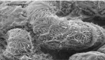
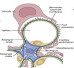
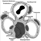
Introductory slide Anatomy of the kidney Histology of the glomerulus (EM)Under the (LM) Scanning EM Under the Under the (EM) Transmission (Cross section) EM Under the (LM)
Normal Kidney Function Excretory function: Detoxify blood, removal of toxins, nitrogenous wastes (urea and creatinine), drugs etc. 1 Homeostatic function: Maintain and regulate water balance and osmolality. Maintain and regulate electrolyte balance (Na+, K+, Ca2+, Cl-, Mg2+ ions etc) . Maintain and regulate acid-base balance (pH, H+ and HCO3- ions). 2 3 Endocrine function: Secretes renin from the JGA cells to regulate BP and electrolyte balance. Secretes erythropoietin from the endothelial cells lining the cortical peritubular capillaries to stimulate the bone marrow to produce RBCs. Converts inactive Vit D3 to active 1,25 dihydroxycholecalciferol by alpha one hydroxylase enzyme under the influence of PTH (Parathyroid hormone) increases calcium absorption.
Important terminology is a clinical syndrome associated with fluid, electrolyte, and hormone imbalances and metabolic abnormalities, which develops with deterioration of renal function. due to the accumulation of organic waste products that are normally cleared by the kidneys. uremia means urine in the blood. Uremia can be seen in both chronic kidney disease and acute kidney injury. Uremia Is abnormally high levels of nitrogen- containing compounds (such as urea, creatinine etc) in the blood. It can lead to uremia if not controlled. It is an elevation of blood urea nitrogen (BUN) and serum creatinine levels. Azotemia Urine output is markedly less than 400 ml/24 hours Oliguria Urine output less than 200 ml/24 hours Anuria is when the kidneys fail to adequately filter toxins and waste products from the blood. In renal failure there is decrease in glomerular filtration rate and an elevated serum creatinine level. Renal Failure (Insufficiency) Note: Creatinine clearance or filtration is dependent on the glomerular filtration rate (GFR).
Kidney biopsy The tissue obtained is used to make slides for: Light microscopy (LM) : to study the histology in renal cortex & medulla. Immunofluorescence (IF) : study is to detect: the presence of immunoglobulins (IgA, IgG, IgM) and complements (C3 and C1q) in the glomerular mesangium or in the wall of the glomerular capillary loops. Electron microscopy (EM) (ultrastructural) : study is to detect the presence or absence of: effacement of the epithelial cell (podocytes)foot processes. electron dense immune complex deposits If deposits are present then to identify the location of the deposits in the glomeruli (mesangial/paramesangial, subepithelial, subendothelial).
Renal failure classification Based on Duration Chronic Acute Sudden onset. Rapid reduction in urine output. Usually reversible. Gradually progressive with nephron loss. Usually not reversible. Based on Etiology 1- Pre-Renal 3- Post-Renal 2- Renal (55-60%) The renal tubular and glomerular function are normal. Here the GFR is decreased due to reduced renal perfusion. (35-40%) (5-10%) Occurs when obstruction in the urinary tract below the kidneys causes waste to build up in the kidneys. Is due to diseases of the kidney itself (which is associated with release of renal afferent vasoconstrictors). Based in the urine output Oliguric Non-oliguric Anuric Urine output less than 400ml/day Urine output greater than 400ml/day Urine output less than 100ml/day NOTE: sometimes more than 70% of renal function can be lost before it is clinically noticeable.
Acute kidney injury (previously called acute renal failure [ARF]) Definition AKI is a syndrome defined by a sudden loss of renal function over several hours to days resulting in the accumulation of nitrogenous compounds such as urea and creatinine. It is characterized by: Note: the contra st mediu Accumulation of nitrogenous waste products. Rapid rise in serum creatinine m/dye used in variou s radiological tests can occasi AKI. onally induce Fluid Oliguria: the urine output is markedly decreased (usually it is less than 400 ml/day) Sudden decrease in GFR (hours to days) imbalance; electrolyte imbalance; acid-base disturbance; and mineral disorders. Pathogenesis Of Acute Kidney Injury Note:In AKI if there is reduction in renal blood flow/ renal perfusion decrease in glomerular filtration rate (this is the common pathologic pathway for AKI regardless of the cause). The decreased GFR activation of renin-angiotensin-aldosterone system. These physiologic changes result in increased sodium reabsorption, increased water retention, increased urinary creatinine concentration, increased urine specific gravity and increased urine osmolality. Acids are retained and HCO 3 is excreted.
Based on Kidney Disease Improving Global Outcomes(KDIGO) AKI is defined by the presence of any one of the following: 1- Pre-Renal azotemia Is the most common cause of acute renal failure (ARF). Initially the body tries to compensate but in severe hypoperfusion GFR is markedly decreased AKI It results from Decreased renal perfusion/hypope rfusion (i.e. decreased effective blood flow to the kidney. The treatment of the cause Restores the renal function. (important to know all of them because it may come as cases) + MCQs Hypovolemia: e.g. hemorrhage, volume depletion (dehydration or GIT fluid loss in vomiting, diarrhea), hypoalbuminemia, diuretics, third space losses (burns, peritonitis, muscle trauma) etc. Pre Renal causes of ARF are: Anything that leads to renal Hypoperfusion: Impaired cardiac function (cardiac failure, myocardial infarction, massive pulmonary embolism). Sepsis, septic shock. Cirrhosis (it can cause renal vasoconstriction kidney injury called hepatorenal syndrome ) Anaphylaxis. Other causes: surgery, NSAIDS and other nephrotoxic drugs etc.
2- Renal causes of AKI (intrinsic) Interstitial & tubulo- interstitial diseases Glomerular diseases Tubular diseases Vascular diseases There is glomerular injury in severe forms of active glomerulonephritis (GN) >> leads to reduction in total filtration area >> reduction in GFR. Examples include: Post infectious GN Rapid progressive crescentic GN Active autoimmune GN Etc. Various forms of vasculitis and emboli lead to reduced renal blood flow due to vascular obstruction or destruction, thus lowering the GFR e.g. Vasculitis Thromboemb olic disease (renal artery/ renal vein thrombosis) Thrombotic microangiopat hies (HUS/ TTP) Malignant hypertension Acute Tubulo- interstitial Nephritis (TIN) e.g. Drug induced TIN Autoimmune TIN Infections TIN Rarely malignant infiltration in interstitium can cause AKI Acute Tubular Injury/ Necrosis It can be ischemic cause or nephrotoxic cause Ischemic: prolonged ischemia to nephrons leads to the tubular injury and necrosis Toxic: toxicity to nephrons leads to tubular injury & necrosis I) Endogenous toxins Pigments and casts: Myoglobinuria (in Rhabdomyolysis) Hemeglobinuria Myeloma casts etc. II) Exogenous toxins Nephrotoxic drugs: Aminoglycosides (antibiotic). Amphotericin B Calcineurin inhibitors (e.g., tacrolimus) etc. Radiograph contrast medium (dyes induced toxicity) 3- Post renal causes of AKI/ ARF Any obstruction to the outflow of urine: Congenital or structural abnormality Benign prostatic hyperplasia (older men) Stones in the urinary tract A tumor in the urinary tract (e.g. ureter, bladder, prostate, urethra). External compression from the outside of the urinary tract (e.g. retroperitoneal fibrosis or tumors like carcinoma of cervix).
Note: Common causes of AKI The kidney biopsy of a patient with AKI can show any of the previously mentioned causes. The common causes are: Tubular cause: acute tubular injury/ acute tubular necrosis Interstitial cause: acute tubulointerstitial nephritis (later lectures) Glomerular cause: Post infectious glomerulonephritis (later lectures) Rapid progressive crescentic glomerulonephritis (later lectures) Acute tubular injury and Acute tubular necrosis In acute tubular injury/ necrosis there is damage to the epithelial cells of the renal tubule and it leads to rapid decline of renal function (i.e. AKI) and presence of granular casts and tubular cells in urine. The damage can be induced by: Ischemia leading to renal ischemic injury: renal ischemia > reduction in GFR > acute tubular injury/ necrosis. The proximal tubule is most vulnerable to ischemia. [renal ischemia can be caused by any condition that causes prolonged persistent hypovolemia or circulatory shock e.g. extensive trauma, burns, hemorrhage, pancreatitis, incompatible blood transfusions, dehydration, septic shock, etc.] 1. 2. Substances toxic to the kidney leading to nephrotoxic injury: here the AKI is caused by direct injury of the tubules by toxins. Causes include: Antibiotics: Aminoglycosides, Tetracyclines, Amphotericin, Cephalosporins etc. Heavy metals: Mercury, Lead, Arsenic, Gold salts, Barium etc. Others: Cisplatin, Doxorubicin, Carbon tetrachloride, Radiographic contrast agents, etc.
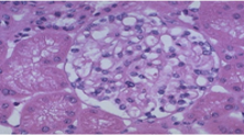
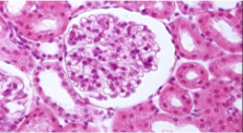
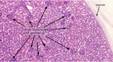
Gross morphology bilaterally enlarged & swollen kidneys (due to edema) Cut surface shows a pale cortex and a dark & congested medulla Histology Ranges from tubular epithelial cell swelling (mild injury) to tubular epithelial cell necrosis (severe injury). The tubular epithelial cells become flattened (so the wall of the tubule becomes thin) and show loss of brush border. In the necrotic tubular epithelial cells there is loss of nucleus and also there is sloughing (falling off) of necrotic cells into the lumen of the tubule. tubular epithelial cell The proximal tubules become dilated and irregular in shape proximal tubules Casts may be present in the lumen of the distal tubules. distal tubules. Interstitium mild edema. Later as healing begins there is regeneration of the tubular epithelial cells Normal tubule Acute tubular injury/ necrosis
Clinical features of AKI Oliguria: Decreased urine output (occasionally urine output remains normal). Typically the urinary output is < 400 ml/day. Defective elimination of metabolic waste, water, electrolytes, and acids from the body. Waste material is accumulated in the body which causes azotemia (> nausea & vomiting), acid-base imbalance (> acidosis) and electrolyte imbalance like hypernatremia and hyperkaliemia (> abnormal heart rhythms, risk of heart failure, weakness and muscle paralysis). Salt and water retention > generalized edema starting with swelling in your legs, ankles or feet. Pulmonary edema (shortness of breath due to extra fluid on the lungs). Other features: hypotension, nephritic syndrome, flank pain, fatigue, uremic encephalopathy, confusion, seizures or coma in severe cases Sometimes acute kidney failure causes no signs or symptoms and is detected through lab tests done for another reason. Treatment Treat the underlying etiology. Dialysis Correction of fluid imbalance. Correction of acidosis and electrolyte imbalance e.g hyperkalemia
Summary - Renal failure (Renal insufficiency): Is when the kidneys fail to adequately filter toxins and waste products from the blood. In renal failure there is decrease in glomerular filtration rate(GFR) and an elevated serum creatinine level. Treatment: treat the cause Renal Failure - - Renal failure classification Based on Duration Acute Chronic - - - Sudden onset Rapid reduction in urine output Usually reversible - - Gradually progressive with nephron loss Usually NOT reversible based on Urine output Oliguric Non-oliguric Anuric Urine output greater than 400cc/24hr Urine output less than 400cc/24hr Urine output less than 100cc/24hr based on Etiology (MCQs) Pre-Renal Renal Post-Renal (55-60%) The renal tubular and glomerular function are normal. Here the GFR is decreased due to reduced renal perfusion. (35-40%) Is due to diseases of the kidney itself (could be vascular or glomerular , tubular, interstitial. ) (5-10%) Occurs when obstruction in the urinary tract below the kidneys causes waste to build up in the kidneys. Tubular Diseases (Acute tubular injury/ Necrosis) Toxicity to nephrons leads to tubular injury & necrosis Reduced renal perfusion due to : Toxicity to nephrons leads to tubular injury & necrosis - Hypovolemia: e.g. hemorrhage, volume depletion (dehydration or GIT fluid loss in vomiting, diarrhea),hypoalbuminemia, diuretics, third space losses (burns, peritonitis, muscle trauma) etc. - Impaired cardiac function (cardiac failure, myocardial infarction, massive pulmonary embolism). Sepsis, septic shock. Cirrhosis (it can cause renal vasoconstriction kidney injury called hepatorenal syndrome ) Anaphylaxis Other causes: surgery, NSAIDS and other nephrotoxic drugs etc. prolonged ischemia to nephrons leads to tubular injury and necrosis. Exogenous toxin Nephrotoxic drugs: - Aminoglycosides (antibiotic). - Amphotericin B - Calcineurin inhibitors (e.g.,tacrolimus) etc. Radiographic contrast medium (dyes induced toxicity) Endogenous toxin Pigments and casts: - Myoglobinuria (in Rhabdomyolysis) - Hemoglobinuria - Myeloma casts etc. -Bilaterally enlarged & swollen kidneys (due to edema). -Cut surface shows a pale cortex and a dark & congested medulla. Gross morphology Ranges from tubular epithelial cell swelling (mild injury) to tubular epithelial cell necrosis (severe injury). The proximal tubules are dilated with thinning of tubular wall. The tubular epithelial cells are flattened and show loss of brush border Hyaline, granular and pigmented casts are present in the lumen of the distal tubules. The interstitium has mild edema Later as healing begins there is subsequent regeneration of the tubular epithelial cells Histological morphology
Quiz Q1) Most common etiology of renal failure: A. B. C. D. Pre-Renal cause Post-Renal cause Renal cause Intrinsic cause Q2) When the urine output is less than 400cc/24hr it s: A. B. C. D. Oliguric Non-oliguric Anuric None of the above Q3) abnormally high levels of nitrogen-containing compounds A. B. C. D. Uraemia Azotemia Anuric Oliguria Q4) Is studying the histology in renal cortex & medulla. A. B. C. D. LM EM IF Ultrasound Q5) Treatment of acute tubular injury and acute tubular necrosis includes all but the following. A. B. C. D. Treat the underlying etiology Dialysis Correction of fluid imbalance. surgery Q6) In typical patient with acute kidney injury which feature is most likely to be present. A. B. C. D. Oliguria ( urinary output is < 400 ml/day) fever coma hypokalemia Q7) Which one of the following cause nephrotoxic injury? A. B. C. D. hemorrhage incompatible blood transfusions Radiographic contrast agents dehydration Answer: Q1/A Q2/A Q3/B Q4/A Q5/D Q6/A Q7/ C
Members Members Team Leaders Maha Alkoryshy Manar Alzahrani Hassan Alabdullatif Team Members Hoor Aloraini Farah alhalafi Shahad Alsalahi Nouf Aldhalaan Jana alhazmi Mayssar Alshobaki Rahaf Almutairi layan Aldoukhi Atheer Alahmari Walaa AlMutawa Fatima Halawi Omar Alkadhi Emad Almutairi Rakan Alromayan Ali Alzahrani Saad Alahmari Malik halees Khalid Alrasheed Nawaf Alturki Pathology442@gmail.com Theme was done by Shatha Almutib


 AKI")
")