Insights into the Plague: Epidemiology, Etiology, and Outbreaks
The Plague, caused by Yersinia pestis, has a chilling history spanning pandemics like the Justinian Plague and the Black Death. Understanding its etiology, family, and pathogenicity is crucial. This deadly disease has had notable outbreaks in India, emphasizing the importance of recognizing its host range and reservoirs.
Download Presentation

Please find below an Image/Link to download the presentation.
The content on the website is provided AS IS for your information and personal use only. It may not be sold, licensed, or shared on other websites without obtaining consent from the author. Download presentation by click this link. If you encounter any issues during the download, it is possible that the publisher has removed the file from their server.
E N D
Presentation Transcript
Plague (Metazoonoses type I) Also K/as Mad rat disease Black death Mahamari Pestilential fever Buboes Pest "Black death" inspired one of the most enduring nursery rhymes in the English language, Ring a Ring O'Roses, a pocket full of posies / Ashes, ashes (or ah-tishoo ah-tishoo), we all fall down .
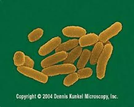
Etiology Yersinia pestis Family: Enterobacteriaceae A gram negative, Non-spore forming, Non-motile, Coccobacillus looks like "safety pin" (i.e. bipolar) (Staining: methylene blue stain or Giemsa's stain or Wayson's stain) Forms capsule: In living tissues Not very resistance to physical & chemical agents
Etiology Coagulase a protein secreted Yersinia pestis Clot the blood Coagulase is inactive at high temperatures: Cessation of plague transmission during very hot weather Pathogenicity is determined by: Fractions (F1), V, W, endotoxin & exodoxin (cardiotoxin) F1 is a capsular heat labile protein (used in serological tests) F1, V & W fractions: Make resistant to phagocytosis Cardiotoxin: lethal to mice & rats
Epidemiology: Pandemics The first pandemic: Justinian s Plaque Started in 542 AD in Egypt Spread to Asia & Europe 100 million deaths The second pandemic: Black Death Started in 1347-1352 AD from Jaffa Spread to China, India & Europe 25 million victims Justinian the Roman Emperor The third pandemic: Started in 1894Yunnan in China Spread to India and to Europe, Asia & Africa Killed more than 12 million people in India & China alone in the period from 1898-1918
Outbreaks: In India Human plague In 1966: Kolar district (Maharastra) In 1994 (August to September): two outbreaks Beed district (Maharashtra) & Surat (Gujarat) Outbreaks suspected to be of plague were reported from Bihar & HP Sylvatic foci of the disease are located in the Deccan plateau, Himalayas & the watersheds of Vindhya, Bhanier & Maikal ranges (MP) foothills of
Epidemiology: Host range & reservoir Primary host: Rodents R. norvegicus Bandicota R. rattus Reservoir of infection: Rat fleas Gerbil (Xenopsylla cheopis) Rodents Fleas may remain infected for months R. rattus Other host: cats, pigs, cattle, sheep, goat, horse, deer etc. Dogs: No overt clinical signs Sentinel animals
Source & Transmission In Animals In rats : By the bite of infected flea Camel: Infection by ingestion of contaminated hay Cats: Feeding on infected rodents Y. pestis: enter in to the body through skin, conjunctiva, oral route & inhalation route Contact with infected rat fleas (X. cheopis) or rodents Pulmonary form spread by airborne or droplet infection Human infections From non-rodent species Direct contact with infected tissues By scratch or bite injuries Handling of infected animals Man to man transmission is mainly air-borne
Source & Transmission The main routes of infection in man are: Domestic rat-rat flea-man (bubonic plague) Wild rodent-flea/ contact-man Wild rodent-flea-domestic rodent-flea-man Man-human flea-man Man-man by droplet (pneumonic plague) By handling of infected rodents Ingestion of contaminated animal tissue Bites of infected cats Bites of ticks, lice and bed bugs Use of clothes infected with flea In India vector of the disease three species of Rat flea: X. cheopis, X. astia & X. brasiliensis The gerbil flea: Nosapsyllus nilgeriensis
Manner in which fleas transmit plague Flea feeds on Y. pestis infected blood Coagulase by Y. pestis Y. Pestis enters flea s midgut & multiplies logarithmically Clump of Y. pestis forms in the midgut, blocking fleas foregut "Blocked flea condition". During next meal, blood cannot enter the midgut & flea gets very hungry Flea bites vigorously & regurgitates the contents of its midgut into the next wound
Importance of flea blockage (A) Unblocked, uninfected flea (B) blocked, infected flea on the right http://www.asm.org/ASM/files/CCLIBRARYFILES/FILENAME/0000000467/nw20030086p.pdf Experiments indicate that only blocked fleas effectively transmit plague to mammals
Disease in animal Primarily effects: Animal of order Rodentia (Effect both wild & domestic rodents) Lesser degree: Rabbits & hairs Infection: Acute, Chronic & Inapparent Domestic rats: more susceptible, Rattus rattus die in large number during to epizootic Acute cases: Haemorrhages with buboes & spleenomegaly, without other internal lesions Sub acute cases: Buboes (caseous), & Punctiform necrotic foci are found in spleen, liver, lung In dogs: Illness is self-limiting, In cats: Severe Often fatal infection, characterized by formation of abscess, lymphadenitis, lethargy, & fever. Kittens: Secondary pneumonia
Disease in man Incubation period: 2-6 days The Three clinical forms: Bubonic plague Septicemic plague Pneumonic plague All three clinical forms start with: Fever, chills cephalgia, nausea, Generalized pain, diarrhoea or constipation, Toxemia, shock, arterial hypotension, Rapid pulse, anxiety, staggering gait, Slurred speech, mental confusion, & prostration
Disease in man Bubonic plague: The most common form Incubation period: Few hrs -12 d (2-7 d after flea bite) Small vesicle at the site of flea bite Symptoms: Fever Swollen, tender lymph nodes, "Buboes". The bubo: 1 to 2 cm Extremely painful & surrounded area become odematous More common: Inguinal, groin & axial region Inguinal buboes: the pain in abdomen, vomiting & diarrhoea, which may be bloody Fatality rate: 25% to 60% (in untreated cases)
Disease in man Systemic plague / Septicemic plague: The bubonic form spreads into the blood Onset of disease: rapid (death in 2 days) Case fatality rate: 100% Develops nervous & cerebral symptoms Symptoms are: Epistaxis Cutaneous petechiae Hematuria Involuntary bowel movements
Disease in man Pneumonic plague: The incubation period : 3 to 5 days The most serious form Primary: by direct inhalation of bacteria Secondary: Derived from bubonic/ Systemic form Symptoms: High fever, chills & often severe headache Cough develops in 24 hours The sputum: Clear at first, Later foamy/haemorrhagic Rapid progressive pneumonia (no pleurisy) Death occurs in 2 days Mortality may exceed 50% Complication: Meningitis
Disease in man Pestis minor: It is a mild form of plague Usually occurs: in endemic area Symptoms are: swollen lymph nodes, fever, headache & exhaustion, which subside within a week Plague is also called the "black death" because disseminated intravascular coagulation takes place & areas of skin undergo necrosis
Diagnosis History & clinical presentation: Bubos, subcutaneous & generalized congestion, granular liver, congested spleen & pleural effusion Demonstration of plague bacilli: Impression smears of aspirates from buboes or blood or sputum Safety pin like morphology Wayson staining (Bipolar stainning) Gram staining
Diagnosis Isolation of organism: On cefsulodin-Irgasan-novobiocin (CIN) agar , blood agar On CIN agar On blood agar Smooth, round opaque, irregular edges, raised center, flat periphery (Fried egg appearance) Grey white translucent colony after 24 h
Diagnosis FAT: Antibodies against F1 antigen Serology: CIE, ELISA, CFT, PHA, Dot-ELISA Molecular diagnosis: PCR Animal inoculation Rapid diagnostic Kit:
Treatment & Control Treatment: Streptomycin chloramphenicol is effective Continuous surveillance: Sentinel animal programmes should be used in endemic areas Prompt diagnosis and medical care Chemoprophylaxis to the exposed group with tetracycline or Rodent control By use of rodenticides like warfarin, zinc phosphate Fumigation with carbon disulphide, SO2, methyl bromide Proper disposal of garbage Proper storage of food grains Trapping Elimination of fleas: BHC as 3% dust or 3% malathion
Treatment & Control Disinfection: Masks, gowns and gloves should be worn when handling cats suspected to be infected. All contaminated surfaces with sputum, discharges & dead rats by sanitation Vaccination: Two doses of formalin killed whole bacteria vaccine are given at interval of 7 to 14 days. Immunity starts one week after vaccination Confers immunity for 6 months Isolation of plague affected people must be done
