Understanding Shock: Causes, Symptoms, and Stages

Shock is a critical condition caused by an imbalance between cellular oxygen supply and demand, leading to organ dysfunction. Compensatory responses include increased heart rate, vasoconstriction, and hormonal adjustments. Recognizable features of shock include tachycardia, hypotension, cool clammy skin, and mental status changes. Lab values show increased lactate, metabolic acidosis, and abnormal organ function markers. Shock progresses through stages of compensated, decompensated, and irreversible, with prompt recognition and treatment crucial for preventing multi-organ failure and death.

Download Presentation
Please find below an Image/Link to download the presentation.
The content on the website is provided AS IS for your information and personal use only. It may not be sold, licensed, or shared on other websites without obtaining consent from the author. Download presentation by click this link. If you encounter any issues during the download, it is possible that the publisher has removed the file from their server.
Presentation Transcript
SHOCK DR . RATI PRABHA ASSOCIATE PROFESSOR KGMU
Definition Shock is the clinical condition of organ dysfunction resulting from an imbalance between cellular oxygen supply and demand. Imbalance between cellular oxygen supply and demand: decreased supply, increased demand, or inadequate oxygen utilization
This leads to anaerobic metabolism, production of lactate, cell damage, organ dysfunction. If not treated immediately causes multiorgan failure and death.
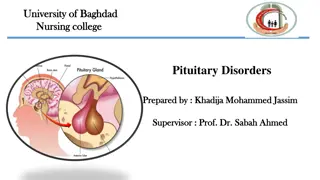
COMPENSATORY RESPONSES Decreased MAP stimulate baroreceptor reflex by sympathetic stimulation causes increase HR and stroke volume. Adrenal medulla releases adrenaline and noradrenaline, causes peripheral vasoconstriction, leads to SVR and MAP. If shock persists(late response), renin is release, RAS is activated causing systemic vasoconstriction. Aldosterone causes sodium and water reabsorption and increase plasma and increase MAP and CO. ADH from posterior pituitary gland causes sodium and water reabsorption
Features Tachycardia, weak and thready pulse Hypotension Tachypnea/dyspnea and shallow breathing Skin: cool, clammy, mottled skin except distributive shock capillary refilling time (normal<3 seconds) Oliguria (<0.5 ml/kg/hr) Mental state changes seen in advanced and decompensated shock. ST elevation.
Increased lactate Decrease pH ( metabolic acidosis) Increased BUN and S. creatinine Increased ALT and AST Increased troponin and proBNP Decreased SvO2 (except septic shock)
Stages Compensated Shock (pre-shock) Decompensated Shock Irreversible Shock
Compensated shock (pre-shock): Initial Phase. Physiological responses counteract the initial insult and attempt to reestablish adequate perfusion and oxygen delivery No obvious signs of organ dysfunction-difficult to recognize E.g. mild increase in lactate/creatinine But treatment is easy. E.g:-10% body blood volume loss in hemorrhagic shock.
Decompensated shock Organ dysfunction starts, and the patient's compensatory response to the insult is overwhelmed. Organ failure starts in this phase. Present with symptomatic tachycardia, dsypnea, restlessness, diaphoresis, metabolic acidosis, hypotension, oliguria and cool, clammy skin. E.g: 20 to 25% reduction in blood volume in hypovolemic shock. fall in cardiac index to less than 2.5 l/min/m2in cardiogenic shock.
Irreversible shock Signs are easily recognized Has high mortality Irreversible organ damage and Multi-organ failure Hypotension becomes so severe and refractory to therapy, acidemia further depresses cardiac output and vasoplegia established Anuria and acute renal failure. Pt is obtundated and can lead to coma and death
Pathophysiologic classification Hypovolemic Distributive Cardiogenic Obstructive Acute blood loss GI loss Burns Anaphylaxis Sepsis Neurogenic shock Adrenal crisis Myocardial infarction Arrhythmia Cardiomyopathy Myocarditis Valvular heart disease Tension pneumothorax Pericardial tamponade Pulmonary embolism Aortic dissection Mixed Shock: features of > 1 type. Eg: Cardiac dysfunction in severe sepsis, severe burn
Hemodynamic features: Preload CVP/PCWP Afterload/ SVR Cardiac Output/ Pulse pressure Lactate Urine output SvO2 Cold/clammy extremities Hypovolemic Yes Cardiogenic Yes / Obstructive Yes /N /N Distributive No
Hypovolemic Shock Decreased intravascular volume: Hemodynamics: features: Preload, Cardiac output, Stroke volume, pulse pressure, CVP, SVR Tachycardia hypotension Urine output, Cool, clammy peripheries, weak pulse CRT, increased lactate Internal or external bleeding, Dehydration, or Gastrointestinal/urinary losses Large burn Third space loss Diuretic
Management: identify cause and treat, Fluid and/or blood transfusion Dynamic indices of intravascular fluid deficit (pulse pressure variation, stroke volume variation, and passive leg raising) are preferred for diagnosing and monitoring ongoing fluid resuscitation. Packed blood cell transfusion is needed to keep hemoglobin level >7 gm/dl. Management If fluid resuscitation cannot maintain tissue perfusion, vasopressors (norepinephrine, vasopressin) are added, and a second etiology of shock is searched.
Distributive Shock Mechanism Hemodynamics: Features: Tachycardia Hypotension Urine output, Warm peripheries, CRT normal, increased lactate Results from widespread severe vasodilatation, leading to a condition of relative hypovolemia. Organ perfusion is decreased. Septic and anaphylactic shock afterload/SVR, BP Preload, N/ Cardiac output/Stroke volume, CVP
Septic Shock- Management Septic shock
Anaphylactic shock Severe, life-threatening allergic reaction that can occur after exposure to an allergen eg antibiotics, insect sting, succinylcholine, latex etc. Management: Removal of trigger IV access not present: IM 0.3 to 0.5 mg epinephrine, administered every 5-10 minutes. IV access present: epinephrine boluses, followed by infusion if needed Rapid IV fluid bolus: crystalloid 1-2 liters Adjunctive agents: Antihistaminic Bronchodilators, oxygen
Cardiogenic shock Hemodynami cs: Mechanism Features: Tachycardia/Bradycardia/arrh ythmias Cardiac dysfunction leading to inadequate organ perfusion and dysfunction. Hemodynamics: Cardiac output & Stroke volume, CVP compensatory SVR hypotension Urine output, Hypotension and reduced cardiac index <2.2 L/minute/m2 Cool, clammy peripheries, weak pulse CRT, increased lactate
Obstructive Shock Mechanism Hemodynamics: features: Extracardiac obstruction of blood flow Tension pneumothorax/ pericardial tamponade: blood flow into RA/RV Pulmonary embolism: outflow from RV to pulmonary circulation Tachycardia hypotension Urine output, Cool, clammy peripheries, weak pulse CRT, increased lactate Hemodynamics varies, but cardiac output is always decreased severely.
Management: Tension pneumothorax: Needle decompression and ICD Pericardial tamponade: drainage Pulmonary embolism: anticoagulation, thrombolysis Aortic dissection: surgical repair of aorta


:")