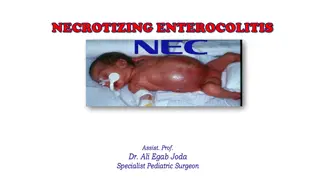
Anesthetic Considerations for Necrotizing Enterocolitis in Neonates

Necrotizing enterocolitis (NEC) is a serious gastrointestinal emergency in neonates, especially those with very low birth weight. Mortality rates are high, and early diagnosis is crucial. Key features, pathophysiology, implications for anesthetic preparation, and diagnostic considerations are discussed in this comprehensive overview. Preterm infants are particularly at risk, with NEC presenting with GI and systemic signs. Radiologic and laboratory findings aid in diagnosis, while prompt management is essential for improving outcomes.
- Neonates
- Necrotizing Enterocolitis
- Anesthetic Considerations
- Preterm Infants
- Gastrointestinal Emergency

Download Presentation

Please find below an Image/Link to download the presentation.
The content on the website is provided AS IS for your information and personal use only. It may not be sold, licensed, or shared on other websites without obtaining consent from the author. Download presentation by click this link. If you encounter any issues during the download, it is possible that the publisher has removed the file from their server.
E N D
Presentation Transcript
Anesthetic Considerations for Necrotizing Enterocolitis Updated 9/2019 Monica Williams, MD Johns Hopkins School of Medicine
Disclosures No relevant financial relationships
Learning Objectives: The learner will be able to identify key features of NEC The learner will be able to describe the pathophysiology of NEC The learner will be able to describe implications of NEC on anesthetic preparation
Background Information Necrotizing enterocolitis (NEC) is the most common gastrointestinal emergency in neonatal intensive care units. True incidence is unknown, but in US proven NEC occurs in 1-3 per 1000 live births. Globally, the reported incidence varies from 2-7% across NICUs. Over 90% of cases are associated with very low birth weight neonates (<1500 g) born <32 weeks. Outcomes Mortality rates range from 15-30% Mortality is inversely related to gestational age and birth weight.
Backgroung information continued NEC in Preterm Infants Incidence of NEC increases in very low birth weight (VLBW) infants (<1500g) and infants born at <32 week gestation. Rates of NEC increased 5-fold for infants <1000g and <28 weeks. NEC in Term Infants 10-15% of NEC cases occur in term infants Studies demonstrate increased incidence in infants receiving non-human milk and have pre-existing illness Associated conditions: sepsis, congenital heart disease, perinatal hypoxia, and fetal growth restriction.
Clinical Presentation NEC can present with both GI and systemic signs. Most common presentation: Sudden change in feeding tolerance Abdominal distension and tenderness Lethargy Respiratory distress Temperature instability (sudden variation and inability to maintain normothermia) Signs of hemodynamic instability Timing of presentation Onset of symptoms varies and may be inversely related to gestational age. Median age of onset of NEC GA less than 26 weeks 23 days GA greater than 31 weeks 11 days
Making the Diagnosis Radiologic findings: Plain x-rays may demonstrate free intraperitoneal air, dilated loops of bowel, ascites, and pneumatosis intestinalis. Ultrasound of abdomen can demonstrate absence of mesenteric blood flow. Contrast enemas are NOT recommended and may cause more harm Laboratory findings (not diagnostic on their own) Thrombocytopenia Metabolic acidosis Coagulopathy Persistantly high levels of C-reactive protein
Abdominal Imaging in NEC Abdominal x-ray of neonate with NEC demonstrating dilated loops of bowels and arrows indicating pneumatosis Santos IG, et al. Radiologia Brasileria 2018.
Making the diagnosis Diagnosis Signs Symptoms Treatment Suspected NEC Abdominal distension and feeding intolerance, heme- positive stool Normal X-rays or mild dilation, no signs of pneumatosis or portal vein gas. Close observation, consider bowel decompression and holding feeds. Consider blood cultures, broad spectrum antibiotics. Medical NEC Same as above but grossly bloody stools X-rays show dilated loops of bowel. Pneumatosis +/- portal vein gas. Bowel decompression and hold feedings for 7-10 days. Close monitoring, blood cultures, antibiotics. Surgical NEC Abdominal distension Free intraperitoneal air on abdominal x- rays Exploratory laparotomy with resection if necessary Surgical NEC Abdominal distension Persistant ileus Placement of drain
Pathophysiology of NEC Probably multifactorial, but not completely understood Preterm infants are predisposed because of immature motility, defenses, absorption and an excessive inflammatory response to luminal microbial stimuli. Based on timing of NEC (at least 8-10 days post partum), inappropriate microbial colonization may also be an important factor. Hypoxia-ischemia may also contribute by modulating microvascular tone A combination of these factors can lead to intestinal necrosis.
Pathophysiology continued Fetal hypoxia and perinatal asphyxia Reduce intestinal motility Watershed areas of ileum and colon are sensitive and are the most commonly affected bowel segments Umbilical artery catheters Can reduce mesenteric blood flow If NEC is suspected, remove catheter Predisposing factors for NEC Prematurity Immature immune system Infection
Medical Management of NEC Bowel rest Bowel decompression Broad spectrum antibiotics Supportive care for hemodynamics, and ventilator support for respiratory compromise
Surgical Management Surgical intervention is necessary when there is bowel perforation, gangrenous bowel, and pneumoperitoneum. Usually the surgery involves resection of the gangrenous bowel, and enterostomy formation. If extremely unstable, can place a peritoneal drain to temporize and optimize care before transporting to an operating room.
Anesthetic Management (Pre-Op) Try to optimize pre-operatively if time allows, and try to correct hypovolemia, metabolic acidosis, coagulopathy, and hypocalcemia. Patients may present on vasopressors, and/or extremely acidotic. Be sure to check labs and ensure blood is available for transfusion. Ensure adequate venous access at least 2 peripheral IVs or 1 peripheral and a central access line.
Anesthetic Management Standard ASA monitoring plus peripheral arterial line Again, ensure adequate venous access In addition to large volume of IV fluids, may need inotropic support (dopamine, epinephrine, or vasopressin) For intermittent bolus dosing, 1mcg/kg/dose of epinephrine can help temporize If possible, follow urine output and look for at least 0.5mL/kg/hr Replace blood loss with blood and fresh frozen plasma
Anesthetic Management Cont Usually these neonates are very sick and already intubated, but if not, an awake intubation or rapid sequence intubation is preferred Avoid nitrous oxide to prevent further bowel distension For maintenance, use air/oxygen mixture to maintain SpO2 around 90%
Anesthetic Management Aggressively avoid hypothermia Very warm OR temperature Radiant heat lamps Warming blankets Warm IV fluids Wrap head and extremities in plastic Warm and humidify gases
Post-operatively Expect to continue ventilation in the NICU Transport in warmed isolette with full monitoring Expect a prolonged ileus and consider central line for TPN and continued inotrope support until sepsis is controlled. Infants that initially respond to medical management or a drain may still eventually need surgical management. Acute complications include infections, disseminated intravascular coagulation, and further metabolic or cardiovascular compromise.
Conclusions: Have a high suspicion for NEC especially for the low birth weight preterm infants. Not every patient with NEC will need a laparotomy, a drain or medical management are options for the correct patient population. Intraoperatively, expect dramatically high fluid requirements, and consider inotropic support. Worry about hypothermia, and aggressively try to prevent. Continue ventilation and cardiovascular support post-operatively
References: 1. Pierro A. The surgical management of necrotizing enterocolitis. Early Hum Dev. 2005 Jan; 81(1): 79-85. Sodhi P, Fiset P. Necrotizing enterocolitis. Continuing education in Anaethesia. 2012; 12 (1): 1-4. Kliegman R. Neonatal necrotizing enterocolitis: bridging the basic science with the clinical disease. J pediatric 1990: 117: 833-835. Hillier S. Neonatal anesthesia. Semin Pediatr Surg. 2004 Aug; 13 (3): 142-151. Kim JH. Neonatal necrotizing enterocolitis: Clinical features and diagnosis. UpToDate. Updated June 21, 2019, from https://www.uptodate.com/contents/neonatal-necrotizing- enterocolitis-management#H7 Neu J. Necrotizing Enterocolitis. N Engl J Med. 2011 Jan 20; 364: 255- 64. 2. 3. 4. 5. 6.


")