Comprehensive Guide to Management of Fractures in Animals
This detailed guide covers the diagnosis and surgical treatment of humeral diaphyseal fractures, supracodylar fractures, and radial and ulnar fractures in animals. It includes information on immobilization techniques, diagnostic imaging, and surgical options for optimal management of bone injuries in veterinary practice.

Download Presentation

Please find below an Image/Link to download the presentation.
The content on the website is provided AS IS for your information and personal use only. It may not be sold, licensed, or shared on other websites without obtaining consent from the author. Download presentation by click this link. If you encounter any issues during the download, it is possible that the publisher has removed the file from their server.
E N D
Presentation Transcript
INTERNAL IMMOBILIZATION OF FRACTURE
HUMERAL FRACTURE HUMERAL DIAPHYSEAL FRACTURES SUPRACONDYLAR FRACTURES
HUMERAL DIAPHYSEAL FRACTURE It results in disruption of continuity of diaphyseal cortical bone Cause : high velocity injuries e.g. Motor vehicle accidents, gunshot injuries, blunt trauma Radial nerve injury may occur with fractures involving the distal humerus HUMERAL DIAPHYSEAL FRACTURES
DIAGNOSIS History Physical examination : Non weight bearing, varying degree of limb swelling, pain and crepitus on manipulation, animal drag the limb while walking Diagnostic Imaging : General anaesthesia is required for proper positioning of animal to obtain best quality radiographs Craniocaudal view Lateral view
SURGICAL TREATMENT Pre-operative management :- Before surgery, a spica splint can be applied to increase patient comfort and protect soft tissue from further injury induced by bone fragments Operative management :- Intramedullary pinning Orthopaedic wire Interlocking nails IM pins plus external skeletal fixation Bone plates
SUPRACONDYLAR FRACTURES These are usually transverse or short oblique fractures Comminuted fracture with multiple small fragments can also be noticed Transverse or short oblique fractures require rotational and bending support Comminuted fracture require axial, rotational and bending support SUPRACONDYLAR FRACTURES
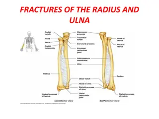
RADIAL AND ULNAR FRACTURE Most often seen in young animals and is frequently comminuted Can occur anywhere in the shaft of bone Metaphyseal & Epiphyseal fractures are common in young animals Avulsion fracture of radius
DIAGNOSIS History Physical examination : Palpation of limb reveals swelling , Pain and crepitation Reluctant to move the limb Diagnostic imaging :craniocaudal and lateral radiographs of affected radius and ulna are required to assess the extent of bone and soft tissue injury
SURGICAL TREATMENT If the fracture is at the distal third of the radius, full limb cast upto the elbow joint provide satisfactory result Internal fixation alongwith some form of external support is usually effective Transfixation can be used except in cases of proximal radial fracture For mid shaft fractures, transfixation along with modified Thomas splint provides satisfactory result Both conventional and compression plating can be used for repair of Radial fracture
FEMORAL FRACTURES FEMORAL DIAPHYSEAL AND SUPRACONDYLAR FRACTURES FEMORAL METAPHYSEAL AND ARTICULAR FRACTURES
. High velocity injuries are the most common type of trauma that causes femoral fractures Most injuries result from Motor vehicle accidents, gunshot injuries, blunt trauma Coxofemoral luxation may occur concurrently with femur fractures Observation of pelvic girdle symmetry and gental rectal palpation help determine the presence of pelvic fracture
DIAGNOSIS History Physical examination : Non weight bearing lameness, Swelling over scapula, crepitation on palpation Diagnostic imaging : Radiographs of femur should include Lateral view Caudocranial view
SURGICAL TECHNIQUES For femoral diaphyseal fracture A. Application of intramedullary pins B. Placement of interlocking nails C. Application of external skeletal fixation D. Application of bone plates and screws C A D B
For metaphyseal fractures A. Stabilization of femoral neck fracture with cancellous lag screw B. Stabilization of femoral neck fracture with triangulated krischner wires
For intertrochanteric fractures A. Stabilization of a Unicondylar fracture with a lag screw B. Stabilization of a Bicondylar fracture with a lag screw & Steinmann pin A B
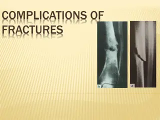
Complications of fracture and repair A fracture and its repair may lead to number of complication which the course of healing of injured area Use of various immobilization devices may cause pressure sores, atrophy of muscles and joint stiffness Main complications related to fracture healing are :- Malunion Delayed union , Non union Avascular necrosis Shortening of bones Infection
CONT. Nerve injury : Radial, Obtrutor, Suprascapular and Peroneal Nerves are more prone to injury Injury to blood vessels : damage to the major blood vessels may occur due to direct trauma Injury to viscera : In fracture of ribs, injury to lungs, heart, diaphragm, spleen and liver may occur In pelvic fracture , urinary bladder and rectum are more prone to injury
Failure of implants Excessive wear and tear of plaster cast, Bending of splints Bending and loosening of plates, screws and intramedullary pins
Malunion (Angular deformity) Healing of the bone fragments in a abnormal position. The causes of malunion are: Faulty reduction. Inadequate immobilization. Failure of fixation of the fracture.
Delayed union / Non-union Delayed union is prolongation of fracture healing. Non union is cessation of fracture healing. The cause are: More gap between the fractured fragments. Loss of blood supply Damage to the surrounding soft tissues Compound fracture or Infection Incomplete fixation Overstripping of periosteum Functional disuse. Early weight bearing.
CONT. Radiographic Signs of Delayed Union Both fractured ends are far apart. Presence of very little callus. Radiographic Signs of Non-union No callus formation. Sclerosis of bone ends. Gap between the bone fragments. Medullary canal is completely closed in each fragment.


")