Distal Radius Fracture: Diagnosis, Management, and Complications
Distal radius fractures, particularly Colles fractures, are common injuries, often affecting elderly women due to osteoporosis. These fractures typically result from a fall onto an outstretched hand, causing dorsal and sometimes radial displacement of the distal radius fragment. Clinical features include the characteristic dinner fork deformity and wrist tenderness. Diagnosis is aided by X-ray imaging to confirm the fracture type. Treatment involves plaster slab immobilization for undisplaced fractures and reduction under anesthesia for displaced fractures, followed by immobilization with a dorsal slab. Early and correct diagnosis, along with appropriate management, are crucial to prevent debilitating complications.

Download Presentation

Please find below an Image/Link to download the presentation.
The content on the website is provided AS IS for your information and personal use only. It may not be sold, licensed, or shared on other websites without obtaining consent from the author. Download presentation by click this link. If you encounter any issues during the download, it is possible that the publisher has removed the file from their server.
E N D
Presentation Transcript
Fracture of the Distal radius Fracture of the Distal radius Dr. Alaa Abdulhussein Dawood Assistant professor Orthopedic Surgery Medical College ,Basrah University
objectives To learn about these very common injuries. To be able to reach a correct ,early diagnosis, and offer a good early management . To be able to prevent important, and serious complications that may lead to crippling or permanent disability.
Colles fracture (Abraham Colles described in 1814) @ Most common fracture in orthopedic practice. @Usual victim is elderly women ( osteoporosis) @It is extraarticular fracture of distal radius . MECHANISM : Fall on the out stretched hand, fracture is at the corticocacellous junction .
Transverse fracture 2.5 cm from distal radius. Distal fragment shifted &tilted dorsally ,also shifted radially.
Clinical features : Typical dinner fork deformity; broad wrist, local tenderness painful wrist movement. look for other injury. Examine for :neurovascular injury- median nerve injury (common)
X-ray: PA ,Lateral &oplique veiw Transverse fracture ; the distal fragment is shifted & tilted dorsally , also shifted radially. Sometimes impacted or comminuted . Often associated with fracture ulnar styloid .
treatment Undisplaced fracture : plaster slab for 4-6 wks Displaced fracture : 1)Reduction : under G.A , or sedation,disimpaction by traction on the hand, the wrist in palmar flexion, ulnar deviation , the forearm pronated, digital pressure on the distal fragment toward palmar side to correct the deformity. Reduction is confirmed by x-ray.
2)Immobilization :below elbow dorsal slab up to metacarpal neck with the wrist fixed in slight palmar flexion& ulnar devation. 3)After care : > Elevation to watch for swelling(examine the fingers for swelling or cyanosis ) >complete pop done once swelling subsided . >follow the patient weekly, by x-ray >redispalacement is not uncommon ,if it occurs, repeat closed reduction. >if alignment is satisfactory ,change the cast after 3 weeks &apply a new cast for further 3 wks. >Encourage shoulder, elbow &finger exercises as early as possible. 4) Surgery : by internal fixation or external fixation in unstable,impacted or comminuted fractures .
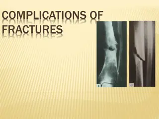
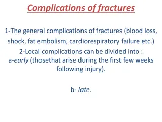
Complications : Early :1)circulatory embarrassment due to tight plaster. 2)Nerve injury: median nerve compression, carpal tunnel syndrome (rare). 3)Reflex sympathetic dystrophy(sudek atrophy) Late : 1)Malunion : common 2)Delayed union or nonunion of ulnar styloid 3)Joint stiffness : of wrist & shoulder is common . 4)Tendon rupture : of extensor pollicis longus tendon .
Smiths fracture (Reversed Colles ) described a similar fracture about 20 years later Uncommon , the distal fragment is displaced anteriorly , Cause : Fall on the dorsum of the hand >Clinical features : Typical deformity : Garden spade deformity
X-ray: PA ,Lateral& oplique (PA) veiw:Transverse fracture of the distal radius Lateral view : the distal fragment is displaced &tilted volarly .
Treatment : Displaced fractures (stable) : !)Reduction :Under G.A ,sedation ,or L.A traction , supination &extension of the wrist. 2)immobilzation : above elbow POP ,forearm supinated & wrist dorsiflexed for 6 weeks . Displaced (unstable) : 1)closed reduction with percutaneous pinning 2) open reduction &fixation by a plate &screws
Fracture of the distal radius in children Fall o the outstretched hand with the wrist in extension ,the distal fragment is displaced dorsally (Juvenile colle s) Sometimes the wrist is in flexion and the distal fragement is displaced anteriorly The fracture is through the distal radial physis or in the metaphysis of one or both bones. Metaphyseal fracture are often incomplete or green stick . http://upload.wikimedia.org/wikipedia/en/7/7d/Salter_Harris_1_demo.jpg
Clinical features : pain, swelling of the wrist ,sometimes there is typical dinner fork deformity . X-ray :accurate diagnosis is made on the x-ray appearance : 1)Physeal fractures: according to salter harris classification usually type 1or 2. Type 5 is unusual .
http://upload.wikimedia.org/wikipedia/en/7/7d/Salter_Harris_1_demo.jpghttp://upload.wikimedia.org/wikipedia/en/7/7d/Salter_Harris_1_demo.jpg
Metaphyseal fractures: Appear as buckling of the cortex Or as angulated greenstick fracture Or as complete # with displacement
Treatment Physeal fractures : reduction under G.A , or sedation ,immobilized in above elbow cast , with elbow in 90 D flexion, wrist slightly flexed &ulnar deviated. Cast retained for 4 weeks. * Buckle fracture: 2 Weeks in p.o.p , then 2 weeks of restricted activity .
Greenstick fractures: closed reduction (easy ). Children <10 years up to 30D angulation Children>10 years up to 15 D angulation is Accepted . Above elbow p.o.p , for 2 weeks, then re x-ray if redisplacement , repeat manipulation . Cast removed after 6 weeks. Complete fractures : Difficult to reduce especially if the ulna is intact, Closed reduction done as colles # , full length cast applied with wrist neutral & forearm supinated .reduction is checked by x-ray , which is repeated after 2 weeks , cast retained for 6 weeks .If # slips, then do closed reduction &percutaneous K wire fixation
Complications : Early : forearm swelling threatened compartmental syndrome . Late : 1)Malunion :uncommon in children<10years because of remodelling power. 2) Radio-ulnar discrepancy :(physeal fracture , especially type 5) premature fusion of radial epiphysis ,leads to subluxation of distal radio ulnar joint.
Radio-carpal fractures: 1)Fracture radial styloid (chauffer fracture ) : forced radial deviation of the wrist after a fall , or when starting handle kick-back . X-ray : transverse fracture line , TRT : closed reduction , wrist held in ulnar deviation by plaster slab , if failed , the fragment is fixed by percutaneous k-wire or screw .
2)Fracture- subluxation(Barton fracture) Types: Volar & Dorsal Barton . Volar Barton (anterior marginal fracture) : oblique fracture through the wrist joint, the distal fragment from anterior articular surface , displaced anteriorly carrying the carpus with it .
TRT : unstable fracture , usually redisplace after closed reduction ,so open reduction& internal fixation by small ant. Plate is recommended
Dorsal Barton(posterior marginal fracture) Oblique fracture through distal radius , the fracture line pass through the wrist joint , Distal fragment from dorsal articular lip , displaced upward &backward carrying the carpus with it.
TRT : 1)try closed reduction& cast for 6 weeks , 2)IF failed;do closed reduction & percutaneous pinning , or open reduction and internal fixation by plate & screws .
3)Comminuted intra articular fractures (young adults) .High energy injury, poor outcome will result unless joint congruity, fracture alignment & length are restored , and movement started early .
TRT : Reduction seldom possible , fracture is unstable & it is difficult to hold the reduction Try closed reduction &above elbow cast ,take serial x-ray for follow-up. If failure : ORIF , or by External fixation(ligamentotaxis)+bone graft
Complications of radiocarpal fractures I. Associated injuries to the carpus II. Redisplacement III. Carpal instability IV. Secondary osteoarthritis
Questions What are the steps in the diagnosis of trauma to wrist due to fall on the outstretched hand. What are the dangerous complications you want to avoid in patients sustaining these injuries? How to avoid the above complications?


")


Immobilization :below elbow dorsal slab up to metacarpal")

")
")


Fracture- subluxation(Barton fracture)")

")
try closed reduction& cast for 6 weeks ,")
Comminuted intra articular fractures")