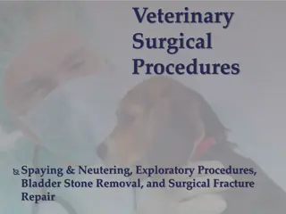
Methods of Internal Immobilization for Fracture Repair

Various methods of internal immobilization for fracture repair in animals include intramedullary pinning and nailing, Rush pins, cross pinning, screws, wires, and plate fixation. These methods provide stability and alignment for bone fractures, with success rates depending on the size and weight of the animal. Pins can be inserted through retrograde or normograde routes, offering different levels of rotational stability. Specific devices like Rush pins and Steinmann pins are used for different types of fractures and bone locations.

Uploaded on Jul 23, 2024 | 0 Views
Download Presentation

Please find below an Image/Link to download the presentation.
The content on the website is provided AS IS for your information and personal use only. It may not be sold, licensed, or shared on other websites without obtaining consent from the author. Download presentation by click this link. If you encounter any issues during the download, it is possible that the publisher has removed the file from their server.
E N D
Presentation Transcript
INTERNAL IMMOBILIZATION OF FRACTURE
INTERNAL IMMOBILIZATION Intramedullary pinning and nailing Rush pins Cross pinning Wires Screws Transfixation Hanging pin cast Plate fixation Steinmann pins
INTRAMEDULLARY PINNING AND NAILING Sound and economical method of internal fixation Steinmann pins, Kuntscher nail (K-nail) is used for repair of long bone fractures in large as well as in small animal A pin should provide 3 point fixation
CONT. Pin provides axial alignment and stability, but little rotational stability A pin should cover approx. 60-70% of diameter of medullary cavity Fracture of Radius, Tibia, Humerus and Femur can be repaired by this method Success rate of this method in large animals depends upon the size and weight of animal Two or more pins (stack pinning) can be used in adult animals especially in femur and humerus Failure are related to mechanical factors such as Pin migration Bending loosening
CONT. Pin is inserted in the medullary cavity by using a simple hand driven chuck Pin can be inserted via 2 routes :- Retrograde : within the fracture site Normograde : from one end of the bone Normograde Retrograde
RUSH PIN Used to treat fractures of the distal femur and supracondylar and diaphyseal fractures of tibia A rush pin is tempered round intramedullary device It has hooked end that is used to drive and seat the pin into the bone Other end is tapered which bounces off the inner cortex as it is inserted into the medullary cavity of bone Elastic bending nature of pins produces a spring like action to provide rigid fixation Generally inserted from distal end of bone Placement of rush pin
CROSS PINNING Useful in compound subarticular fractures of long bones Especially tibia, metacarpus and metatarsus Can also be used to repair fracture of mandible
WIRES In orthopaedic surgery different types of wire are used The rigid kirschner wire, kirschner wire The flexible orthopaedic wire Suture wire. Orthopaedic wire Suture wire
ORTHOPAEDIC WIRE It is a monofilament soft and flexible wire. Full circlage: The wire should be fixed perpendicular to the long axis of the bone and the knot must be twisted down snugly. Hemicirclage: Hemiciriclage wiring is effective in reinforcing longitudinal cracks in the cortex & prevent rotation and overriding of oblique fracture fragments Tension band wiring or figure of 8 wiring :used in conjunction with Steinmann pins to achieve stable internal fixation by opposing the pull of muscular attachment on bone. Kirschner wire: Kischner wires are used for temporary fixation of fragments, tension band osteosynthesis and intramedullary fixation in small bones. Full circlage Hemicirclage Tension band wiring
SCREWS Cortical These screws are full threaded and used where cortical bone predominates The inter-fragmentary compression is accomplished by drilling a long gliding hole (oversized hole) in the near cortex and a smaller threaded hole in the far cortex. Cancellous The screws are partially threaded used in cancellous bone e.g. fracture of the olecranon, slab, fractures of the metacarpus and metatarsus, condylar fractures and longitudinal fracture of the phalanx can be fixed with screw. Oblique fracture of long bones can also be fixed by application of screw in combination with internal or external support. Cortical Screws Cancellous Screws
TRANSFIXATION Most useful for treatment of diaphyseal fracture of the radius and tibia. A minimum of two intramedullary pins in each fractured fragment are inserted transversely The protruding ends of intramedullary pins are fixed in position by connecting external bars and protected with caps. The pins are connected by one or more connecting bars. The assembly should be removed only after complete union of fractured fragments Transfixation pins
COMPLICATION: Soft tissue infection, Bone necrosis, Periosteal reaction around the transverse pins. Pathological fracture may occur at the point of insertion of transverse pins.
HANGING PIN CAST Only one pin is inserted tissue transversely through the proximal fragment . This technique has the advantage of preventing rotation of the fractured bone & downward slipping of the plaster cast.
PLATE FIXATION Plate provide axial compression, counteract rotational forces, can effectively immobilize oblique and comminuted fracture. Plate classification:( according to the function) Buttress Plate Compression plate: Static compression (a transverse or short oblique fracture can be best treated by compression plate Neutralization Plate: Splinting and lag screw fixation (comminuted fracture are anatomically reconstructed). Buttress Plate: Splinting or bridging a fracture area with buttress of the main fragments. Neutralization Plate
DYNAMIC COMPRESSION PLATE (DCP) DCP is used for compression and stabilization of a fracture. Compression is achieved by tightening the screw inserted in a specially designed hole in the plate. There are three sizes of DCP used in small animal surgery (2.7 mm, 3.5mm and 4.5 mm). At least 3 cortical screw on each side of the fracture fragment should be used.
SCAPULAR FRACTURE May occur through the body, spine, acromion, neck, supraglenoid tuberosity and glenoid cavity Uncommon in dogs and cats bcoz large muscles surrounding scapula protect it from direct injury Common concurrent injuries include : Thoracic injuries Pulmonary contusions Rib fracture Nerve injury
DIAGNOSIS History Physical examination : Non weight bearing lameness, Swelling over scapula, crepitation on palpation Diagnostic imaging : Radiographs of scapula should include Lateral view Caudocranial view
SURGICAL TREATMENT Fixation systems applicable for scapular fracture includes oPlates and screws , oOrthopaedic wires and oKirschner wires Use of Plating (B) and Orthopaedic wires (D) to repair transverse fracture Use of crossed krischner wire