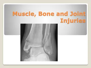
Understanding Fractures: Classification and Factors

A fracture is a break in bone continuity, classified by causal factors, presence of external wounds, location, morphology, severity, and stability post-reduction. Fracture causes include direct violence, indirect violence, bone diseases, and repeated stress. Fractures can be closed or open, with severity graded as simple, wedge, or complex with varying degrees of complexity. Different bone zones and orientations of fracture lines provide additional details for assessment and treatment.

Download Presentation

Please find below an Image/Link to download the presentation.
The content on the website is provided AS IS for your information and personal use only. It may not be sold, licensed, or shared on other websites without obtaining consent from the author. Download presentation by click this link. If you encounter any issues during the download, it is possible that the publisher has removed the file from their server.
E N D
Presentation Transcript
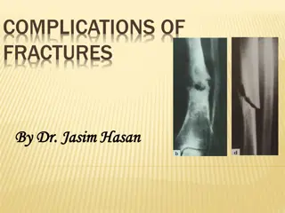
A fracture is a complete or incomplete break in the continuity of bone or cartilage. CLASSIFICATION OF FRACTURES include causal factors; presence of a communicating external wound; location, morphology, and severity of the fracture; and stability of the fracture after axial reduction of the fragments. A. Causal Factors: Direct Violence Applied to Bone. 75% to 80% of all fracturescar accidents or motorized vehicles. Indirect Violence. The force is transmitted through bone or muscle to a distant point where the fracture occurs (e.g., fracture of femoral neck, avulsion of tibial tubercle, fracture of condyles of the humerus or femur).
A. Causal Factors: Direct Violence Applied to Bone. 75% to 80% of all fracturescar accidents or motorized vehicles. Indirect Violence. The force is transmitted through bone or muscle to a distant point where the fracture occurs (e.g., fracture of femoral neck, avulsion of tibial tubercle, fracture of condyles of the humerus or femur). Diseases of Bone. Some bone diseases cause bone destruction or weakening to such a degree that trivial trauma may produce a fracture (e.g., bone neoplasms, nutritional disturbances affecting bone). Repeated Stress. Fatigue fractures in small animals are most frequently encountered in bones of the front or rear foot (e.g., metacarpal or metatarsal bones in the racing greyhound).
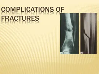
B. Presence of Communicating External Wound: Closed Fracture. The fracture does not communicate to the outside Open Fracture. The fracture site communicates to the outside. These fractures are contaminated or infected, and healing at best may be complicated and delayed.
C. Location, Fracture Morphology, and Severity: Localization of the fracture is provided by; 1. numbering each long bone (1, humerus; 2, radius/ulna; 3, femur; 4, tibia/fibula) and 2. dividing each bone into 1, proximal; 2, shaft; and 3, distal zones. 3. As a measure of severity, each fracture is typed as A, simple; B, wedge; or C, complex. Each grade is further grouped into three degrees of complexity (e.g., A1, A2, A3) depending on the type and extent of bone fragmentation.
Proximal and distal zones may require individual descriptions to accommodate the specific bone morphology.
The orientation of the fracture line relative to the bone s long axis allows the following descriptions: Transverse Fracture. The fracture crosses the bone at an angle of not more than 30 degrees to the long axis of the bone. Oblique Fracture. The fracture describes an angle of greater than 30 degrees to the long axis of the bone. Spiral Fracture. This is a special case of oblique fracture in which the fracture line curves around the diaphysis. Incomplete Fracture. only disrupts one cortex, an incomplete fracture is called a greenstick fracture in young animals because of the bending of the nonfractured cortex. Fissure fractures exhibit fine cracks that penetrate the cortex in a linear or spiral direction. In skeletally immature animals the periosteum is usually left intact. Complete Fracture. A complete fracture describes a single circumferential disruption of the bone.
Multifragmental Fractures. Also known as comminuted fractures, multifragmental fractures have one or more completely separated fragments of intermediate size. These fractures can be further described as follows: Wedge fracture. A multifragmental fracture with some contact between the main fragments after reduction. Reducible wedges. Fragments with a length and width larger than one third the bone diameter. After reduction and fixation of the wedge(s) to a main fragment, the result is a simple fracture. Nonreducible wedges. Fragments with a length and width less than one third the bone diameter and that result in a defect between the main fragments after reduction of more than one third the diameter. Multiple or segmental fracture. The bone is broken into three or more segments; the fracture lines do not meet at a common point. This is a special case of a reducible wedge fracture.
Proximal and distal metaphyseal zones require specific nomenclature to describe the wide variety of extraarticular and intraarticular fractures seen in these locations, as follows: Extraarticular Fractures. The articular surface is not fractured but is separated from the diaphysis. These are typically called metaphyseal fractures. In a physeal fracture the fracture-separation occurs at the physeal line or growth plate. This type occurs only in the young, growing animal.
Partial Articular Fractures. Only part of the joint surface is involved, with the remaining portion still attached to the diaphysis. Unicondylar fractures are the most common example Complete Articular Fractures. The joint surface is fractured and completely detached from the diaphysis Humeral T or Y fractures are representative of this type
The following additional descriptive terms are applied to certain fractures: Impacted Fracture. The bone fragments are driven firmly together. Avulsion Fracture. A fragment of bone, which is the site of insertion of a muscle tendon, or ligament, is detached as a result of a forceful pull.
Stability after Replacement in Normal Anatomical Position Stable Fracture. Fragments interlock and resist shortening forces (e.g., transverse, greenstick, impacted). The primary objective of fixation is to prevent angular and rotational deformity. Unstable Fracture. The fragments do not interlock and thus slide by each other and out of position (e.g., oblique, nonreducible wedges). Fixation is indicated to maintain length and alignment and to prevent rotation.
Score 9 or 10 Fracture transverse or short oblique; type A: 1. Cast/splint 2. IM pins in many, but not all cases; may be combined with interfragmentary wires. 3. Compression plate 4. External fixator, type IA 5. Interlocking nail Score 8 (7) to 9 Fracture long oblique or spiral; type A and B1 one reducible wedge: 1. IM pins/cerclage-hemicerclage wires 2. Neutralization plate 3. External fixator, type I, II (may be combined with cerclage wires/lag screws) 4. Interlocking nail (may be combined with cerclage wires/lag screws) Score 4 (3) to 7 Fracture wedge; type B: 1. Neutralization plate 2. External fixator, type IA double bar or IB, II (may be combined with cerclage wires/lag screws) 3. Interlocking nail (may be combined with cerclage wires/lag screws) Score 1 to 3 Fracture complex; type C: 1. Buttress/bridging plate, or plate and IM pin combination 2. External fixator, type II or III 3. Interlocking nail 2 Fractures: Classification, Diagnosis, and Treatment 145