Understanding Zygomaticomaxillary Complex Fractures
Zygomaticomaxillary complex fractures, commonly referred to as ZMC fractures, are significant facial injuries often caused by trauma. These fractures can involve different parts of the zygoma bone, leading to various classifications based on severity and displacement. Proper diagnosis and appropriate management are crucial to avoid complications and restore normal facial function. Various terminologies and classifications help in identifying and treating ZMC fractures effectively.

Uploaded on Apr 18, 2024 | 7 Views
Download Presentation
Please find below an Image/Link to download the presentation.
The content on the website is provided AS IS for your information and personal use only. It may not be sold, licensed, or shared on other websites without obtaining consent from the author. Download presentation by click this link. If you encounter any issues during the download, it is possible that the publisher has removed the file from their server.
Presentation Transcript
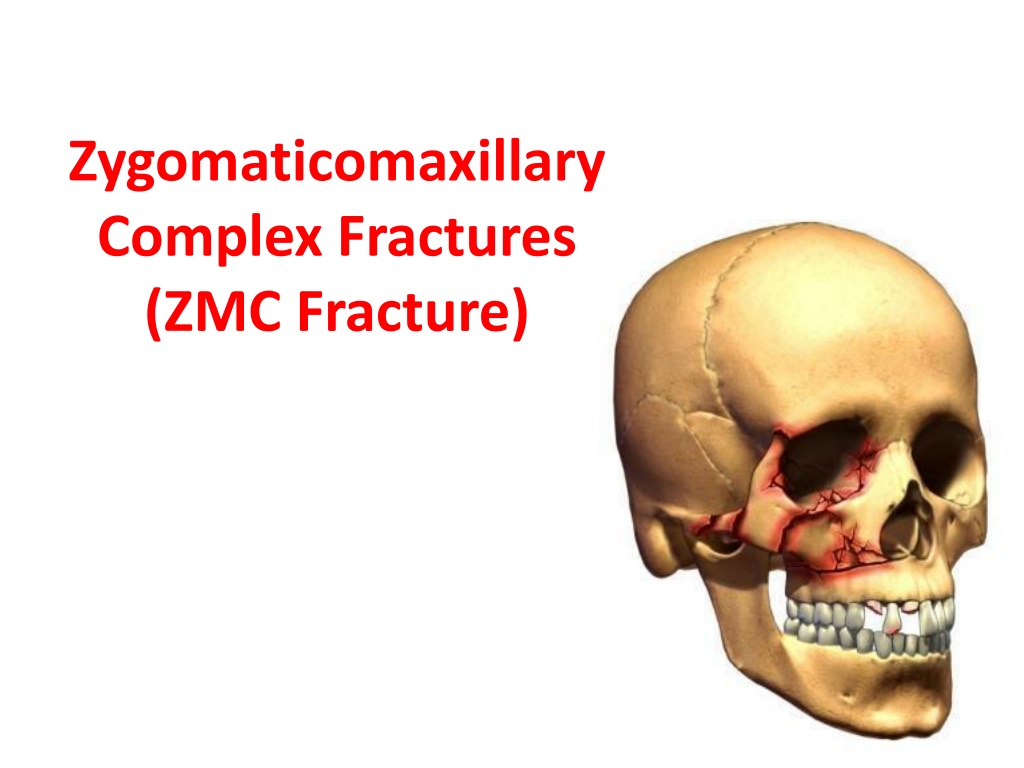
Zygomaticomaxillary Complex Fractures (ZMC Fracture)
ANATOMY OF ZYGOMA BONE Pyramid Shape of Zygoma (Tetrapodal configuration)
Introduction Zygomatic fractures are common facial injuries, representing the second mostin frequency after nasal fracture. The high incidence of these fracture -the zygoma s prominent position. Male:female ratio - 4 : 1 Peak incidence - second and third decades of life 80% - motor vehicle accident. The left zygoma is most commonly affected
DIFFERENT TERMINOLOGIES 1) Zygomatic fracture. 2) Malar fracture. 3) Zygomaticomaxillary complex fracture. 4) Zygomaticomaxillary compound fracture. 5) Zygomaticoorbital fracture. 6) Zygomatic complex fracture. 7) Trimalar fracture. 8) Tripod fracture (Ungley and Suggit, 1944). 9) Tetrapod fracture. (FONSECA et al. FRACTURES OF ZYGOMATIC COMPLEX AND ARCH. ORAL & MAXILLOFACIAL TRAUMA: WB SAUNDERS. 3rd ed. Vol 1)
Classification Knight & North Group I fractures: In these patients fracture lines in zygoma could be seen only in imaging. There is absolutely no displacement. These patients could ideally be managed conservatively by observation and by asking the patient to eat soft diet. Group II fractures: This group includes isolated fractures of the archof zygoma. These patients present with trismus and cosmetic deformities. Group III fractures: This include unrotated fractures involving body of zygoma. Group IV fractures: This involves medially rotated fractures of body of zygoma
Group V fractures: This involves laterally rotated fractures of body of zygoma. This type of fracture is very unstable and cannot be managed by closed reduction. Open reduction will have to be resorted to. Group VI fractures: This is complex fracture. It has multiple fracture lines over the body of zygoma. This condition is difficult to manage by closed reduction. Open reduction and microplate fixation is indicated in these patients. This type of fracture should not be managed by closed reduction alone because the presence of oedema / haematoma would mask the cosmetic deformity giving an impression that reduction has occurred. After reduction of oedema and followed by the action of masseter the fractured fragment may distract making the cosmetic deformity well noticeable
ZINGGetal classification(1992) Type A: Incomplete zygomatic fracture. Isolated fractures involving only one zygomatic pillar: Type A1: Isolated ZAfracture Type A2: Lateral orbital wall fracture Type A3: Infra orbital Rimfracture
Type B: Complete monofragment zygomatic fracture (tetrapod fracture). All 4 pillars of the zygomatic boneare fractured
Type C: Multifragment zygomatic fracture. Same as type B, but with fragmentation, including the body of the zygoma
Manson's (1990) classification of fracture zygoma 1. Low energy injury 2. Medium energy injury 3. High energy injury
SIGNS AND SYMPTOMS Periorbital ecchymosis and edema
Flattening of malar prominence (70% to 86% cases, Larsen et al 1978 and Ellis et al 1985)
TRISMUS ( ONE-THIRD OF THE CASES AND 45 % CASES IN ARCH FRACTURES)
Abnormal nerve sensibility (50% to 90%) (Larsen et al, 1978 and Ellis et al, 1985)
EPISTAXIS (30% to 50%) (Weisenbaugh JM, 1970 and Ellis et al 1985)
Subconjunctival ecchymoses (50% to 70%) (Weisenbaugh JM, 1970 and Ellis et al 1985)
CREPITATION FROM AIR EMPHYSEMA (DISAPPEARS IN 2-4 DAYS SPONTANEOUSLY)
Superior Orbital Fissure Syndrome Superior orbital fissure syndrome, also known as Rochon-Duvigneaud's syndrome, is a neurological disorder that results if the superior orbital fissure is fractured. Involvement of the cranial nerves that pass through the superior orbital fissure may lead to diplopia, paralysis of extraocular muscles, exophthalmos, and ptosis. Blindness or loss of vision indicates involvement of the orbital apex, which is more serious, requiring urgent surgical intervention. Typically, if blindness is present with superior orbital syndrome, it is called orbital apex syndrome
IMAGING CT scans allow detailed examination of the orbit walls and all ZMC buttresses and buttress-related sutures in axial, coronal, and sagittal views . There is also the possibility of producing digital and even stereolithographic 3-dimensional reconstructions of the scan for easier spatial visualization of the sustained injury . The use of ultrasonographic imaging in the diagnosis of craniofacial trauma is increasing. A recent systematic review concluded that the use of diagnostic ultrasonography in maxillofacial fractures, especially fractures involving the nasal bone, orbital walls, anterior maxillary wall, and zygomatic complex, is justified on the grounds that the sensitivity and specificity of ultrasonography were considered generally comparable with those of CT. (Adeyemo et al. IJOMS 2011)
GENERAL PRINCIPLES OF TREATMENT No treatment Indirect reductionwith, a. No fixation b. Temporarysupport c. Direct fixation d. Indirectfixation Direct reduction andfixation
NOTREA TMENT Cases with a minimal degree of displacement, which following union, are considered unlikely to result any cosmetic deformity, disturbanceof vision, persistent paraesthesia or impairment of mandibularmovement.
INDIRECTREDUCTION NOFIXATION: Includes procedures which do not involve exposureof the fracturesites. The principle is to disimpact and reduce the fracture by direct application of an instrument, through approachremote from the fractureline. an indirect
The techniques which have been developed for thisoperative approach, are based upon the introduction of an instrument through, a. the temporalfossa, b. the upper buccal sulcus(intraoral), c. the cheek(percutaneous), d. the nose(transantral) e. the eyebrow (lateralbrow)
Temporal fossaapproach: This method was introduced by Gillies et al (1927) for elevation of the zygomaticarch. Incision of (2 cm in length), made 2.5 cm superior and anterior to the helix, within the hairline made above and parallel to the anterior branch of the temporal artery and dissection is carried down to the temporal fascia. This fascia is then incised to expose the temporalis muscle. An instrument is inserted deep to the temporalis fascia and superficial to the temporalis muscle Using a Back-and-Forth Motion the instrument is advanced untilit is medialto thedepressed zygomaticarch.
Firm Upward and outward force to thelifting handle Use of Rowe s zygomatic elevator(1966)
Elevation from eye brow approach: (Dingman & Natwig1964) Theadvantageof thistechniqueisthat thefractureatthe orbital rim is visualizeddirectly. Thefrontozygomaticareaof thelateralorbital rim is exposed by the eyebrowincision. The instrument is inserted to lift thezygoma anteriorly, laterally andsuperiorly. Useful instruments for this purpose are Dingman zygomatic elevator , urethral sound, or even largeKelly hemostat.
Dingmanzygomaticelevatorisplacedalongthetemporalsurfaceof zygomafor anterior , lateral and superiorelevation
Upper buccal sulcus: (keensapproach) Theadvantagesof thistechniquehavebeendiscussed by Balasubramaniam (1967) who considers that less force is required by the intraoral approach than by the extraoral, because the force is exerted where it should be,i.e.,moreatthecentreofthefractured fragment . Access is gained by an incision of about 1cm in length at the reflection of the upper buccal sulcus immediately behind the zygomatic buttress, so that a pointed curved elevator can be passed upwards supraperiosteally to contact the infratemporal surface of thezygomatic bone. This enables upward ,forward and outward pressureto beexerted.
The elevator by Monks issuitable for this purpose. (Taylor monk spattern) A right angle retractor,bone hook, large Kelly Hemostat, simple dental extraction forceps and a Flat instrument Seldin retractor to followmedial Surface of zyg arch and elevate itlaterally. Quinn in 1977 described a modification which is of valuefor medially displaced fractures of zygomaticarch. This employs a lateral coronoid approach throughan incisionsituatedoverthe anterior borderof ramus.
Percutaneous approach: (Stroymeyer1844) Simplest of all because no soft tissue dissection isnecessary. Several instruments bone hook , carroll Girardscrew( large bone screw ) for elevatingzygomas. This method consists of inserting a hook through the soft tissueofthemalararea atapointjustinferiorand posterior to the prominence of the zygoma so that it engages the infratemporalaspect. Poswillo advises that the exact location of the initial stab wound for insertionis found at the intersection of a perpendicular line dropped from the outer canthus of the eye and a horizontal line extended posteriorly from thealar margin of thenostril.
Carroll-Girard screw elongated cork screw with a T bar handle and threads on its working end. This screw can be threaded into the body of zygoma following placement ofhole Adv control ZMC position in all 3 planes of space. Anterior and lateral traction withbone hook
Intranasal transantral approach: (Lothrops approach1906) Not common inuse. An opening is made into the antrum below the inferior meatus, and a curved urethral sound introduced and manipulated so that its tip lies on the antral aspect of the zygomatic bone. Firm outward and upward pressure is applied to reposition thebone.
ASSESSMENT OFREDUCTION Thesuccessorfailureofreductionwill beobviousforthose who have opened the fracture at three sites. If exposure at three sites has not been performed, the orbital margins are the areas that should be palpated first to determine reduction. If reduction has been satisfactory, these margins will be smooth and continuous. This finding by itself, however, is inadequate verification . Although the zygomaticofrontal suture area provides the strongest pillar of the zygoma, it is oneoftheworstindicatorsofproperreductionofthe entire complex, even when surgically exposed and evaluated directly.
One should also palpate in the maxillary vestibule. If there is any flatness still visible, then zygoma has not been properly elevated. If there is any doubt about proper reduction, exposure is mandatory. In this case, an incision in themaxillary vestibule offers excellent exposure of the zygomaticomaxillary buttress and the infraorbitalrim.
Fixation: 1 Point Fixation 2 Point fixation 3 point fixation 4 point fixation
One point fixation: Indication: Undisplacedfracture. Simple non comminuted zygomatic complexfracture Approach: Zygomaticomaxillary buttress approached through maxillary vestibularapproach.
Two point fixation: Indication: Displaced fracture unstable after reduction Fracture at Frontozygomatic suture, Infraorbital rimand buttress. Approach: Exposure of frontozygomatic suture A 2 point fixation using low profile plate at zygomaticomaxillary buttress or at theand FZ suture
Three point fixation Fixation is done at Frontozygomatic suture, Zygomaticomaxillary buttress and the Infraorbitalrim. Good reduction of these 3 sites mostly reduces the archfracture which is not fixed


")


 classification of fracture")


")
")
")


")


")