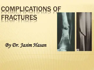
Understanding Different Types of Fractures and Their Characteristics
Explore various types of fractures such as traumatic, pathologic, compressive, and more through detailed medical images and authentic reports. Learn about the severity of soft tissue damage, classifications by renowned professionals, and the significance of assessing soft tissue injuries. Delve deep into descriptions of fractures like open, closed, transverse, oblique, spiral, longitudinal, and their implications on musculoskeletal health. Gain insights into complex fracture patterns, treatment approaches, and the importance of proper diagnosis in managing bone injuries.

Download Presentation

Please find below an Image/Link to download the presentation.
The content on the website is provided AS IS for your information and personal use only. It may not be sold, licensed, or shared on other websites without obtaining consent from the author. Download presentation by click this link. If you encounter any issues during the download, it is possible that the publisher has removed the file from their server.
E N D
Presentation Transcript
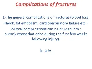
Authentic medical reports De collement = severe damage of soft tissues
Fractura pathologica Myeloma -The plasma membrane of a muscle fiber
Fractura spiralis / longitudinalis
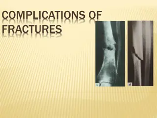
Fractura compressiva / impressiva
Fractura cum dislocatione ad latus ad axim ad longitudinem cum contractione ad longitudinem cum distractione
Authentic reports :1 Fr. aperta TSCHERNE I - open fracture with small skin injury without its contusion - negligible bacterial contamination Profesor Dr. Harald Tscherne (1933), Traumatology Clinic, Hannover: Classification of fractures published in 1982, T. divides fracture into open and closed. The most important is for him the degree of the soft tissues damage.
The acetabulum (cotyloid cavity) is a concave surface of the pelvis. The head of the femur meets with the pelvis at the acetabulum, forming the hip joint.
AO Classification of fractures S 4220 Fractura colli chirurgici humeri l. dx. comminutiva AO 11-C3
Fracture Healing: 1: REPOSITIO = REDUCTIO fragmentorum CLOSED (short /long term)
Fracture Healing: 2: FIXATIO = STABILISATIO fragmentorum PLASTER CAST INTERNAL FIXATION
Fracture Healing: 2: FIXATIO = STABILISATIO fragmentorum INTERNAL FIXATION
Fracture Healing: 2: FIXATIO = STABILISATIO fragmentorum
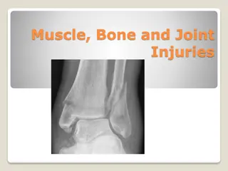
Name the type of fracture B D F A C E
A) Name different bones of the human body B) Write down different types of fractures of named bones
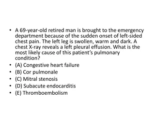
1 A 45-year-old woman presented with a 3-month history of generalized body pains nonresponsive to analgesic agents. Along with low back pain, she had progressive difficulty in getting up from sitting and supine positions and in walking. There was no history of trauma or any medication intake. She is an orthodox believer who wears a black veil outdoors and is completely covered, with little exposure to the sun. An anteroposterior radio- graph of the pelvis showed an undisplaced transverse fracture of the shaft of both femurs. The patient was treated with therapeutic doses of calcium and vitamin D supplements.
2 An 18-year-old slightly intoxicated man was assaulted with a glass bottle on the left parietal region of his head and had a 5- minute loss of consciousness. Two hours after the injury he was presented to a local emergency with severe headache, nausea, and repeated vomiting. Computed tomography of the head revealed a 2.5-cm epidural hematoma in the left parietal region (Panels A and B) underlying a linear nondisplaced skull fracture (Panel C, arrows).
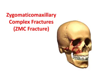
3 A 21-year-old man presented after being struck with a gun on his right lower jaw. Examination revealed displacement of the left half of his mandible with malocclusion on biting (Panel A). Computed tomography showed a fracture of the left mandible and a fracture of the right mandibular body and angle (Panel B). Given the U shape of the mandible, it is common for contralateral fractures to result from major injury. Intravenous analgesics and antibiotics were given; the patient underwent open reduction with internal fixation of his fractures.
Authentic reports :1 De collement = severe damage of soft tissues
Literature Maz nek, J.: Traumatologie orofaci ln oblasti. Praha : Grada, p. 24 http://radiologymasterclass.co.uk http://anthropology.si.edu http://nejm.org (The New England Journal of Medicine)


 is a")


 Name different bones of the human body")