Understanding Prokaryotic and Eukaryotic Chromosome Organization
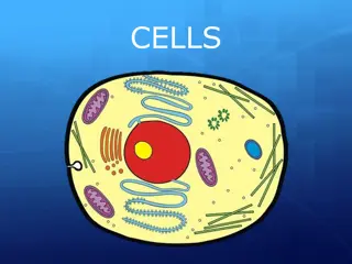
Chromosomes are vital structures in cells, holding genetic material. Prokaryotic cells have a nucleoid containing DNA while eukaryotic cells have DNA enclosed in a nucleus. Proteins like H-NS, HU, FIS, and IHF play crucial roles in maintaining chromosome structure and gene expression. Unlike eukaryotic histones, prokaryotic DNA-binding proteins do not form nucleosomes but use other mechanisms for compaction. Overall, these proteins organize the genome by driving events like DNA bending, bridging, and aggregation.

Download Presentation

Please find below an Image/Link to download the presentation.
The content on the website is provided AS IS for your information and personal use only. It may not be sold, licensed, or shared on other websites without obtaining consent from the author. Download presentation by click this link. If you encounter any issues during the download, it is possible that the publisher has removed the file from their server.
E N D
Presentation Transcript
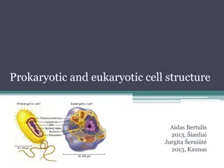
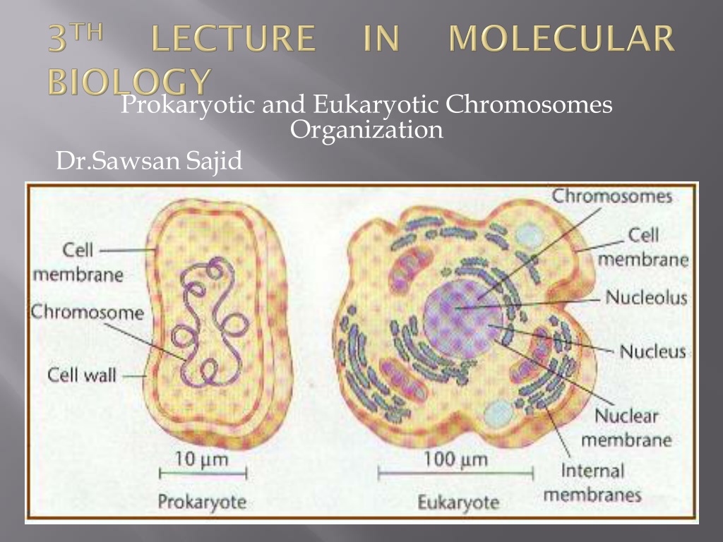
Prokaryotic and Eukaryotic Chromosomes Organization Dr.Sawsan Sajid
- The term chromosome comes from the Greek words for color (chroma) and body (soma). Scientists gave this name to chromosomes because they are cell structures, or bodies, that are strongly stained by some colorful dyes used in research. - Chromosomes are thread-like or supercoiled structures located inside the cell (in cytoplasm of prokaryotes or nucleus of the eukaryotes). Each chromosome is made of protein and a single molecule of deoxyribonucleic acid (DNA). - The nucleoid :(meaning nucleus-like) is an irregularly-shaped region within the cell of a prokaryote that contains all or most of the genetic material. - In contrast to the nucleus of a eukaryotic cell, it is not surrounded by a nuclear prokaryotic organisms generally is a super coiled double-stranded piece of DNA. The length of a genome widely varies, but generally is at least a few million base pairs It is commonly referred to as a prokaryotic chromosome. membrane. The genome circular, of
The chromosome for this bacterium is circular and this is a common arrangement, but there are a number of species with linear chromosomes. If stretched out, this material would be about 1400 m long, an amazing degree of packing is necessary to fit the chromosome inside its tiny host. The genetic material here is composed from 60%DNA the rest is10% protein an large amount of RNA polymerase transcription factor proteins that regulate the expression of genes. These seem not to perform any structural role, but reflect the importance of RNA transcription in the nucleoid in growing cells. and mRNA rRNA,
The nucleoid is composed of DNA in association with a number of DNA-binding proteins (histone-like protein s) that help it maintain its structure. There are four major families of bacterial histone-like proteins: 1- Histone like nucleoid structuring protein(H-NS): It belongs to a family of bacterial proteins that play a role in the formation of nucleoid structure,. It regulates gene expression by binding to AT-rich DNA, which is a common feature of promoters. 2- Heat unstable protein(HU): This protein binds non- specifically to DNA and bends it, the DNA apparently wrapping around the HU protein. 3- Factor for inversion stimulation(FIS): It binds to a loose 15 nucleotide consensus sequence transcription of tRNA and rRNA genes and regulates its own synthesis. 4- Integration host factor(IHF): It facilitates bending of DNA by binding to specific DNA sites. in DNA. it activates
- Proteins associated from histones of eukaryotic nuclei. - In contrast to histones, the DNA-binding proteins of the nucleoid do not form nucleosomes, in which DNA is wrapped around a protein core. Instead, these proteins often use other mechanisms to promote compaction such as DNA looping. The most studied NAPs are HU, H-NS, FIS, and IHF that organize the genome by driving events such as DNA bending, bridging, and aggregation. - These proteins can form clusters and they seem to be involved also in coordinating transcription events, spatially sequestering specific genes and participating in their regulation. or proteins nucleoid proteins or nucleoid- distinct (NAPs) and are
- the chromosome is further folded into 50 or `100 loops(domains) of about 100 kbp. - These domains supercoiled and indeed, even small sections within the same loop can transiently degrees of supercoiling. - the specific supercoiling of a region can affect the ability of the cell to express genes in that region. independently have different
The unique structure of eukaryotes chromosome keep DNA tightly wrapped around spool-like proteins, called histones. Without such packaging, DNA molecules would be too long to fit inside cells. For example, if all of the DNA molecules in a single human cell were unwound from their histones and placed end- to-end, they would stretch 180 cm. Each species of plants and animals has a set number of chromosomes. for example, Humans have 46 chromosomes while a rice plant has 12 and a dog 39.
Basic information about chromatin :it is the major component of the nucleus,the genetic material consist from 50% DNA and protein for each and during interphase this chromatin appeared as uncondensed diffused material look like beads- in string but during metaphase it will arrange to thread-like structure
The beads are called nucleosomes. Each nucleosome is made of DNA wrapped around eight histone proteins that function like a spooland are called a histone octamer . Histones are a family of basic proteins that associate with DNA in the nucleus and help condense it into chromatin. Nuclear DNA does not appear in free linear strands; it is highly condensed and wrapped around histones in order to fit inside of the nucleus and take part in the formation of chromosomes. Histones are basic proteins, and their positive charges allow them to associate with DNA, which is negatively charged. Some histones function as spools for the DNA to wrap around. Each histone octamer is composed of two copies each of the histone proteins H2A, H2B, H3, and H4. The chain of nucleosomes is then wrapped into a 30 nm spiral called a solenoid, where additional H1 histone proteins are associated with each nucleosome to maintain the chromosome structure. each nucleosome attached with followed one by linker DNA ( 20-60 BP) thus total DNA warp around it which will be protected from digestion with micrococcal endonuclease. The fifth histone H1 usually exist out side the core (in the binding region between nucleosome and another)
All four of the core histones amino acid sequences contain between 20 and 25% of lysine and arginine. Molecular size for the core protein ranges between 11.4 KD and 15.4 KD making them relatively small yet highly positively charged proteins allowing them to closely associate with negatively charged DNA, for H1 Histone it is relatively larger (MW :21 KD)and percentage of basic amino acid is 30.5% Five major families exist: H1/H5, H2A, H2B, H3 and H4.Histones H2A, H2B, H3 and H4 are known as the core histones, while histones H1 and H5 are known as the linker histones. of histones
Out side the nucleosome structure there are other type of proteins called non histone protein .usually they are Acidic rather than basic protein doesn't play roles in DNA packing they are the only protein that remain after removing histone during division and clear during metaphase. much larger in molecular weight (more than 30 KD) and irregular heterochromatin rather regular.
First level twisting or super coiling of DNA molecules Second level warping of DNA around histons Formation of folds or zig zag by HI histone and the linker (other benefits of linker create elasticity and flexibility to chromatin beside binding two adjacent nucleosome ) Formation of (30 nm) fibers and salenoid model by collecting each 6 nucleosome together
Telomeres are repetitive stretches of DNA located at the ends of linear chromosomes. They protect the ends of chromosomes. In many types of cells, telomeres lose a bit of their DNA every time a cell divides. Eventually, when all of the telomere DNA is gone, the cell cannot replicate and dies. White blood cells and other cell types with the capacity to divide very frequently have a special enzyme that prevents their chromosomes from losing their telomeres. Because they retain their telomeres, such cells generally live longer than other cells. Telomeres also play a role in cancer. The chromosomes of malignant cells usually do not lose their telomeres, helping to fuel the uncontrolled growth that makes cancer so devastating.