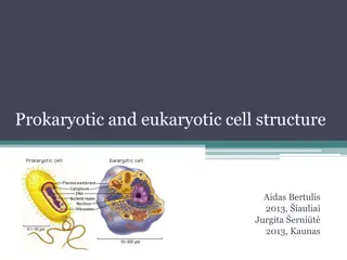
Prokaryotic and Eukaryotic Cells: A Comparative Overview
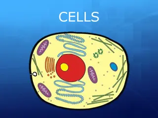
Prokaryotic cells are simpler and lack membrane-bound organelles, reproducing through binary fission. Eukaryotic cells are more complex, larger, with a nucleus enclosed in a nuclear envelope. They have various organelles and a cell wall. The plasma membrane defines cell boundaries, regulating the passage of substances. Both cell types have distinct characteristics and functions within the sub-cellular fractionation process.

Download Presentation

Please find below an Image/Link to download the presentation.
The content on the website is provided AS IS for your information and personal use only. It may not be sold, licensed, or shared on other websites without obtaining consent from the author. Download presentation by click this link. If you encounter any issues during the download, it is possible that the publisher has removed the file from their server.
E N D
Presentation Transcript
PROKARYOTIC and EUKARYOTIC CELLS SUB-CELLULAR FRACTIONATION
The term prokaryote is derived from the Greek word pro, (meaning: before) and karyon (meaning: kernel). It translates to before nuclei. Prokaryotic cells are comparatively smaller and much simpler than eukaryotic cells. It does not possess membrane-bound cell organelles such as a nucleus. Reproduction happens through the process of binary fission. Cells contain special structures called mesosomes which assist in cellular respiration. Most prokaryotes also contain plasmids, which contains small, circular pieces of DNA. To help with locomotion, flagella are present, though, pilus can also serve as an aid for locomotion.
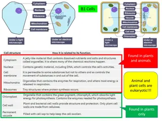
The term Eukaryotes is derived from the Greek word eu , (meaning: good) and karyon (meaning: kernel), therefore, translating to good or true nuclei. Eukaryotes are more complex and much larger than the prokaryotes. Eukaryotes possess a cell wall, which supports and protects the plasma membrane. The cell is surrounded by the plasma membrane and it controls the entry and exit of certain substances. There are also other cell organelles that perform various other functions and these include ribosomes, lysosomes, Golgi bodies, cytoplasm, chromosomes, vacuoles and centrosomes.
The plasma membrane defines the periphery of the cell, separating its contents from the surroundings. It is composed of lipid and protein molecules that form a thin, tough, pliable, hydrophobic barrier around the cell. The membrane is a barrier to the free passage of inorganic ions and most other charged or polar compounds. The internal volume bounded by the plasma membrane, the cytoplasm, is composed of an aqueous solution, the cytosol, and a variety of suspended particles with specific functions. The cytosol is a highly concentrated solution containing enzymes and the RNA molecules. All cells have, for at least some part of their life, either a nucleus or a nucleoid. The nucleoid, in bacteria, is not separated from the cytoplasm by a membrane; the nucleus, in higher organisms, consists of nuclear material en- closed within a double membrane, the nuclear envelope. Cells with nuclear envelopes are called eukaryotes (Greek eu, true, and karyon, nucleus ); those without nuclear envelopes bacterial cells are prokaryotes (Greek pro, before ).
Separation of cellular compartments from one another is an important step for studying a specific intracellular structure or organelle or protein. Assess possible associations between these macromolecular structures. Subcellular fractionation uses one or more of the properties of each compartment, such as buoyant density, surface charge density, size and shape, and is mainly based on differential centrifugation in media of high viscosity at 4 C. Media used for differential centrifugation are mainly sucrose, mannitol, glycerol, Ficoll 400 (a polymer of sucrose). Gel filtration, affinity chromatography, electrophoresis or selective density-shift perturbation can also be used.
ISOLATION OF CELLS OR TISSUE: Initially subcellular fractionation studies were only carried out with tissue, preferentially liver, but also kidney. The liver is built of hepatocytes, endothelial cells, Kupffer cells, and stellate cells. To be sure about the origin of the organelles, one would have to separate the cells first. 1.
2. Homogenisation is the most critical step in every procedure. The aim is to open most of the cells, but keeping the organelles as intact as possible. The best compromise will be the final conditions. 3. Separation of organelles into fractions is usually carried out by centrifugation, although other techniques have been used (2-phase extraction, electrophoresis, immunoisolation for instance). 4. Biochemical analysis: Analysis of marker enzymes specific for certain organelles, or markers introduced to organelles, for instance by endocytosis.
Animal and plant cells are typically 5 to 100 m in diameter, while many bacteria are only 1 to 2 m long. Mycoplasma, sometimes classified as bacteria are 300 nm in diameter, while a single bacterial ribosome is about 20 nm in its longest dimension. The general rule is: the larger the cell, the easier to open less force is needed. The homogenization is usually carried out in an isotonic buffer with sucrose and neutral pH. The isotonisity avoids osmotic effects to influence the integrity of the organelles. Hypotonic medium is used in some cases to aid the rupture of the cell membrane, because it induces swelling of the cells that reduces the force needed.