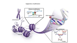
Understanding Plasmids: DNA Molecules Free of Chromosome
Plasmids are DNA molecules existing free of the chromosome in a cell. They can be circular or linear and carry genes beneficial to the host. Plasmids replicate from unique origins and regulate copy numbers through various mechanisms. Different replication mechanisms, such as theta and RC, are used, and plasmids can replicate by more than one mechanism. The copy number of a plasmid is regulated differently based on the type. Various plasmids have unique characteristics and partitioning mechanisms to ensure equal distribution during cell division.

Download Presentation

Please find below an Image/Link to download the presentation.
The content on the website is provided AS IS for your information and personal use only. It may not be sold, licensed, or shared on other websites without obtaining consent from the author. Download presentation by click this link. If you encounter any issues during the download, it is possible that the publisher has removed the file from their server.
E N D
Presentation Transcript
Plasmids are DNA molecules that exist free of the chromosome in the cell. Most plasmids are circular, but some are linear. The sizes of plasmids range from a few thousand base pairs to almost the length of the chromosome itself. Probably the best distinguishing characteristic of a plasmid is that it has a typical plasmid origin of replication with an adjacent gene for a Rep protein rather than a chromosome origin with an oriC gene, along with a dnaA gene and other genes typical of the chromosomal origin of replication.
Plasmids usually carry genes for proteins that are necessary or beneficial to the host under some situations but are not conditions. * Plasmids replicate from a unique origin of replication, or oriV characteristics of a given plasmid derive from this ori region. They include the mechanism of replication, copy number control, partitioning, and incompatibility. If other genes are added to or deleted from the plasmid, it will retain most of its original characteristics, provided that the ori region remains. essential under all region. Many of the
Many plasmids replicate by a theta mechanism, with replication forks moving from a unique origin with leading and lagging template strands, much like circular bacterial chromosomes. Others use an RC mechanism, similar to that used to replicate some phage DNAs and during bacterial conjugation. In RC replication, the plasmid is cut at a unique site, and the Rep protein remains attached to the 5 end at the cut through one of its tyrosines. The free 3 end is used as a primer to replicate around the circle, displacing one of the strands. When the circle is complete,the 5 phosphate is transferred from the Rep protein to the 3 hydroxyl to form a single-stranded circle. A strand complementary to the single-stranded circle is made, using a different origin, and then the host ligase joins the ends to form two double-stranded circular DNAs. Linear plasmids replicate by more than one mechanism. Some have hairpin ends and replicate from an internal origin around the ends to form dimeric circles that are then processed to form two linear plasmids. Others have a terminal protein at both 5 ends and extensive inverted-repeat sequences at their ends.
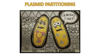
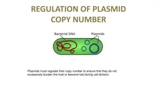
The copy number of a plasmid is the number of copies of the plasmid per cell immediately after cell division. Different types of plasmids use different mechanisms to regulate their initiation of replication and therefore their copy numbers. Some plasmids use small complementary RNAs (ctRNAs). In ColE1-derived plasmids, the ctRNA, called RNA I, interferes with the processing of the primer for leading-strand replication, called RNA II. In other cases, including the R1, ColIB-P9, and pT181 plasmids, the ctRNA interferes with the expression of the Rep protein required to initiate plasmid DNA replication. Iteron plasmids regulate their copy numbers by two interacting mechanisms. They control the amount of the Rep protein required to initiate plasmid replication, and the Rep protein also couples plasmids through their iteron sequences. Some plasmids have a special partitioning mechanism to ensure that each daughter cell gets one copy of the plasmid as the cells divide. These partitioning systems usually consist of two genes for proteins and a cis-acting centromere like site.
If two plasmids cannot stably coexist in the cells of a culture, they are said to be incompatible or to be members of the same Inc group. They can be incompatible if they have the same copy number control system or the same partitioning functions. The host range for replication of a plasmid is defined as all the different organisms in which the plasmid can replicate. Some plasmids have very broad host ranges and can replicate in a wide variety of bacteria. Others have very narrow host ranges and can replicate only in very closely related bacteria. Many plasmids have been engineered for use as cloning vectors. They make particularly desirable cloning vectors for some applications because they do not kill the host, can be small, and are easy to isolate. Some plasmids can carry large amounts of DNA and are used to make BACs for eukaryotic- genome sequencing. Plasmids have been adapted to be used to express cloned genes in bacteria, to do gene replacements in the chromosome.
Plasmid Structure Most plasmids are circular, although linear plasmid also exist. In a circular plasmid, all of the nucleotides in each strand are joined to another nucleotide on each side by covalent bonds to form continuous strands that are wrapped around each other. Such DNAs are said to be covalently closed circular and, because there are no ends to rotate, the plasmid can maintain supercoiled stress. nonsupercoiled DNA strands exhibit a helical periodicity of about 10.5 bp, as predicted from the Watson-Crick double- helical structure of DNA. In contrast, in a DNA that is supercoiled, the two strands are wrapped around each other either more or less often than once in about 10.5 bp. If they are wrapped around each other more often than once every 10.5 bp, the DNA is positively supercoiled; if they are wrapped around each other less often, the DNAis negatively supercoiled.
Like circular plasmid DNAs are usually negatively supercoiled Because DNA is stiff. This makes the plasmid more compact, so that it migrates more quickly in an agarose gel . In the cell, the DNA wraps around proteins, which relieves some of the stress. The remaining stress facilitates some reactions plasmid, such as separation of the two DNA strands for replication or transcription the chromosome, covalently closed involving the
PURIFYING PLASMIDS The structure of plasmids can be used to purify them away from chromosomal DNA in the cell. Bacterial cells are usually lysed with a combination of a strong base (sodium hydroxide), the detergent sodium dodecyl sulfate SDS) to solubilize the membranes, and a component to denature proteins. Following this treatment, a highsalt (potassium acetate) solution is added. Under these conditions, the chromosome precipitates with the cell debris and SDS upon centrifugation, while the small supercoiled plasmids remain in solution. Following these steps, a variety of procedures are used to further purify the plasmid DNA from the proteins and to concentrate the plasmid preparation. Historically, a solution of phenol and chloroform was added to the solution to separate the DNA from the proteins. After mixing of the aqueous lysate with phenol and chloroform, the components quickly separate into phases, similar in concept to oil and water. Plasmid DNA is separated from the proteins because the polar DNA molecules remain in the aqueous phase while the proteins remain at the interface of the solutions.
Following extraction, plasmid DNA is concentrated using precipitation with a salt and alcohol solution and finally resuspended in water or a buffered solution. For convenience today, commercially columns are usually used instead of separation using the phenol-chloroform solution. The plasmid solution is added to a resin matrix in a column, which binds DNA. An alcohol solution is used to wash away the residual proteins and salts from the column, and the plasmid DNA is finally eluted from the column using water or a buffered solution. The isolated plasmid DNA can then be visualized on a gel, digested with a restriction endonuclease, or sequenced, among other applications Visual methods for detectinglarge plasmids involve separating them from the chromosome directly by electrophoresis on agarose gels available spin
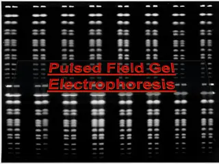
The plasmid, because of its unique size, makes a sharp band on the gel, distinct from that due to chromosomal DNA, which is usually fragmented and so gives a more diffuse band. Also, methods such as pulsed-field gel electrophoresis have been devised to allow the separation of long pieces of DNA based on size Such methods separation of DNA molecules hundreds of thousands of base pairs long and the detection of very large plasmids. have allowed the
Properties of Plasmids Replication DNA molecules that can replicate autonomously in the cell are called replicons. Plasmids, phage DNA, and the chromosomes are all replicons, at least in some types of cells. To be a replicon in a particular type of cell, a DNA molecule must have at least one origin of replication, or ori site, where replication begins . In addition, the cell must contain the proteins that enable replication to initiate at this site. Plasmids encode only a few of the proteins required for their own replication. In fact, many encode only one of the proteins needed for initiation at the ori site.
All polymerases, ligases, primases, helicases, and so on are borrowed from the host. Each type of plasmid replicates by mechanisms, which is determined along with other properties by its ori region The plasmid replication origin is often named oriV for ori vegetative, to distinguish it from oriT, which is the site at which DNA transfer initiates in plasmid conjugation . Most of the evidence for the mechanisms described below came from electron microscope observations of replicating plasmid DNA. of the other required proteins DNA one of two general
THETA REPLICATION Some plasmids begin replication by opening the two strands of DNA at the ori region, creating a structure that looks like the Greek letter hence the name theta replication . In this process, an RNA primer begins replication, which can proceed in one or both directions around the plasmid. In the first case, a single replication fork moves around the molecule until it returns to the origin, and then the two daughter DNAs separate. In the other case (bidirectional replication), two replication forks move out from the ori region, one in either direction, and replication is complete (and the two daughter DNAs separate) when the two forks meet somewhere on the other side of the molecule. The theta mechanism is the most common form of DNA replication, especially in gram- negative bacteria like the proteobacteria. Commonly used plasmids, including ColE1, RK2, and F, as well as the bacteriophage P1, use this type of replication.
ROLLING-CIRCLE REPLICATION Other types of plasmids replicate by very different mechanisms. One type of replication is called rolling- circle (RC) replication because it was first discovered in a type of phage where the template circle seems to roll while a copy of the plasmid is made and processed to unit length. Plasmids that replicate by this mechanism are sometimes called RC plasmids. This type of plasmid is widespread and is found in the largest groups of bacteria, as well as archaea. In an RC plasmid, replication occurs in two stages. In the first stage, the double-stranded circular plasmid DNA replicates to form another double-stranded circular DNA and a single-stranded circular DNA. This stage is analogous to the replication of the DNAof some single- stranded DNA phages and to DNA transfer during plasmid conjugation
In the second stage, the complementary strand is synthesized on the single-stranded DNA to make another double-stranded DNA. First, the Rep protein recognizes and binds to a palindromic sequence that contains the double-strand origin (DSO) on the DNA. Binding of the Rep protein to this sequence might allow the formation of a cruciform structure by base pairing between the inverted-repeated sequences in the cruciform. Once the cruciform forms, the Rep protein can make a nick in the sequence. It is important for the models that the Rep protein is also known to function as a dimer, at least in some plasmids. After the Rep protein has made a break in the DSO sequence, it remains covalently attached to the phosphate at the 5 end of the DNA at the nick through a tyrosine in one copy of the Rep protein in the dimer, as shown.
DNA polymerase III (the replicative polymerase uses the free 3 hydroxyl end at the break as a primer to replicate around the circle, displacing one of the strands. It may use a host helicase to help separate the strands, or the Rep protein itself may have the helicase activity, depending on the plasmid. Once the circle is complete, the 5 phosphate is transferred from the tyrosine on the Rep protein to the 3 hydroxyl on the other end of the displaced strand, producing a single-stranded circular DNA. This phosphotransferase reaction and requires little energy. The same reaction is used to re-form a circular plasmid after conjugational transfer . process is called a
It is less certain what happens to the newly formed double-stranded DNA when the DNA polymerase III has made its way all around the circle and gets back to the site of the DSO.
REPLICATION OF LINEAR PLASMIDS As mentioned above, some plasmids and bacterial chromosomes are linear rather than circular . In general, linear DNAs face two problems in all organisms. One issue with linear DNAs is that the cell must have a way to distinguish the normal DNA ends at the ends of the linear fragments from ends formed when DNA double- strand breaks occur, which would otherwise be lethal to the cell and must quickly be repaired. A second problem with linear plasmids and chromosomes has to do with replicating the lagging-strand template, the strand that ends with a 5 phosphate, all the way to the end of the DNA. This has been called the primer problem because DNA polymerases cannot initiate the synthesis of a new strand of DNA.
Different linear DNAs solve the primer problem in different ways. Some linear plasmids have hairpin ends, with the 5 and 3 ends joined to each other. These plasmids replicate from an internal origin of replication to form dimeric circles composed of two plasmids joined head to tail to form a circle. These dimeric circles are then resolved into individual linear plasmid DNAs with closed hairpins at the ends. The hairpin ends are presumably not recognized as DNA double- strand breaks, because they are not targets for exonucleases in the cell. A completely different mechanism is also used to maintain linear plasmids in some systems. With this mechanism, a special enzyme called a terminal protein attaches to the 5 ends of the plasmid DNA . It is interesting that bacteria with linear plasmids also often have linear chromosomes, and the two DNAs replicate by similar mechanisms.
Functions of the ori Region In most plasmids, the genes for proteins required for replication are located very close to the ori sequences at which their products act. Thus, only a very small region surrounding the plasmid ori site is required for replication. As a consequence, the plasmid still replicates if most of its DNA is removed, provided that the ori region remains and the plasmid DNA is still circular. Smaller plasmids are easier to use as cloning vectors, as discussed below, so often the only part of the original plasmid that remains in a cloning vector is the ori region. In addition, the genes in the ori region often determine many other properties of the plasmid. Therefore, any DNA molecule with the ori region of a particular plasmid will have most of the characteristics of that plasmid.
Not all bacteria have circular chromosomes and plasmids. Some, including Borrelia burgdorferi (the causative agent Streptomyces spp., Rhodococcus fasciens, have linear chromosomes and often multiple linear plasmids. the replication of the ends of linear DNAs presents special problems because DNA polymerases cannot prime their own replication. This means that they cannot replicate all the way to a 3 end in a linear DNA. of Lyme tumefaciens, disease), and A.
Eukaryotic chromosomes, which are linear, solve this problem by having special DNA regions called telomeres at their ends. Most telomeres do not sequences to be synthesized from the template as during normal DNAreplication. Most use an enzyme called telomerase. The Nobel Prizes in physiology and medicine in 2010 went to people who determined how these enzymes work. Telomerase contains an need complementary
Telomeres solve the problem of replicating linear DNAs without losing sequence information. However, bacteria with linear chromosomes are not known to have telomeres made by telomerases and must use different strategies. Two different strategies for dealing with linear chromosomes in bacteria are exemplified in Streptomyces and Borrelia. In both examples, the chromosome replicates from an ori sequence located toward the middle of the chromosome, from where replication occurs bidirectionally using the DnaA initiator protein in a system that is likely similar to those of other bacteria. However, the ways in which the linear ends are maintained in these organisms are very different.
The Streptomyces has inverted-repeat sequences at its ends and a protein, terminal protein (TP), attached to the 5 ends. Replication to the 3 ends of the chromosome is thought to involve both these inverted repeats and TP in a process called patching. After the linear DNA replicates, the 3 end of each DNA remains single stranded, which allows the inverted-repeat sequences to form hairpins. Replication of these hairpins, combined with some sort of slippage, then allows complete replication of the ends by a process that is not completely understood. very large linear chromosome of
The mechanism used by Borrelia to replicate its linear chromosome is very different from that found in Streptomyces spp. and is illustrated in the figure. In Borrelia, the 5 phosphate and 3 OH at each end of its linear chromosome are joined to each other to form hairpins, as shown. When replication initiated in chromosome gets to the ends, the linkage between the 5 phosphate and 3 OH hairpins forms a dimerized chromosome, with two copies of the chromosome forming a circle containing two copies of the chromosome linked end to end . An enzyme called telomere resolvase protein (ResT) the center of the
then recreates the original hairpin ends from these double circles by making a staggered break in the two strands where the original ends were and then rejoining the 3 end of one strand to the 5 end of the other strand to form a hairpin. The ResT enzyme works somewhat topoisomerases and tyrosine recombinases in that the breaking and rejoining process goes through a 3 phosphoryltyrosine intermediate, where the 3 phosphate end is covalently joined to a tyrosine before it is joined to the 5 hydroxyl end. like some
Bacteria that have linear chromosomes may contain linear or circular plasmids, and the same is true for bacteria with circular chromosomes. This suggests that minimal cellular adaptation may be needed to maintain a chromosome in either the linear or circular form. Experiments in E. coli have provided a clever and clear way to help address this question. A lysogenic Siphoviridae bacteriophage called N15 that was found in an isolate of E. coli uses the same mechanism for maintaining its linear genome as that found in Borrelia, with a cis-acting telomere region called tos and a trans-acting resolvase protein, TelN. (lambda-like)