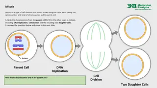
Understanding Chromatin Organization and Chromosome Structure in Molecular Biology
Chromosomes are the carriers of genetic information in cells, containing genes made of DNA. Chromatin, composed of DNA wrapped around histone proteins, plays a crucial role in organizing genetic material. Humans have 23 pairs of chromosomes, and the Human Genome Project aims to map the human genome. The unique structure of chromosomes keeps DNA tightly packed and allows for gene expression and cell functions.

Download Presentation

Please find below an Image/Link to download the presentation.
The content on the website is provided AS IS for your information and personal use only. It may not be sold, licensed, or shared on other websites without obtaining consent from the author. Download presentation by click this link. If you encounter any issues during the download, it is possible that the publisher has removed the file from their server.
E N D
Presentation Transcript
7thlecture in molecular biology Chromatin organization and chromosome structure
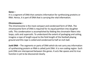
General identification 1-Chromosome: Components in a cell that contain genetic information. Each numerous genes. Chromosomes occur in pairs: one obtained from the mother; the other from the father. chromosome contains 2-Gene:The fundamental physical and functional unit of heredity. A gene is an ordered sequence of nucleotides located in a particular position on a particular chromosome that functional product (ie, a protein or RNA molecule). encodes a specific 3- DNA: The material inside the nucleus of cells that carries genetic information. The scientific name for DNA is deoxyribonucleic acid
Basic structure of chromosome What is a chromosome? Chromosomes are thread-like structures located inside the nucleus of animal and plant cells. Each chromosome is made of protein and a single molecule of deoxyribonucleic acid (DNA). The term chromosome comes from the Greek words for color (chroma) and body (soma). Scientists gave this name to chromosomes because they are cell structures, or bodies, that are strongly stained by some colorful dyes used in research. The unique structure of chromosomes keeps DNA tightly wrapped around spool-like proteins, called histones. Without such packaging, DNA molecules would be too long to fit inside cells. For example, if all of the DNA molecules in a single human cell were unwound from their histones and placed end-to-end, they would stretch 6 feet the condensed nucleoprotein structure is called chromatin . How many chromosomes do humans have? Humans have 23 pairs of chromosomes, for a total of 46 chromosomes .In fact, each species of plants and animals has a set number of chromosomes. A fruit fly, for example, has four pairs of chromosomes, while a rice plant has 12 and a dog, 39 . Human Genome Project: The Genome Project (HGP) work to map the human genome down to the nucleotide (or base pair) level and to identify all the genes present in it.. a
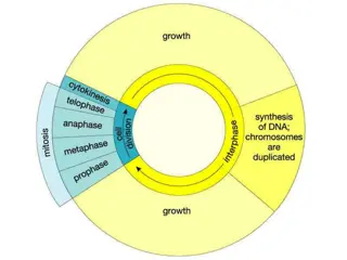
Basic information about chromatin :it is the major component of the nucleus,the genetic material consist from 50% DNA and protein for each and during interphase this chromatin appeared as uncondensed diffused material look like beads- in string ( )but during metaphase it will arrange to thread-like structur
The beads are called nucleosomes. Each nucleosome is made of DNA wrapped around eight histone proteins that function like a spool and are called a histone octamer .Histones are a family of basic proteins that associate with DNA in the nucleus and help condense it into chromatin. Nuclear DNA does not appear in free linear strands; it is highly condensed and wrapped around histones in order to fit inside of the nucleus and take part in the formation of chromosomes.Histones are basic proteins cose they are rich with basic amino acid including arginine and lysine , and their positive charges allow them to associate with DNA, which is negatively charged. Some histones function as spools for the thread-like DNA to wrap around. Each histone octamer is composed of two copies each of the histone proteins H2A, H2B, H3, and H4. The chain of nucleosomes is then wrapped into a 30 nm spiral called a solenoid, where additional H1 histone proteins are associated with each nucleosome to maintain the chromosome structure.
The nucleosome forms by the association of two copies each of the four core histones, H2A, H2B, H3 and H4 forming the histone octamer and ~147 base pairs of DNA. The DNA adopts a flat left handed superhelix with ~1.65 (1 and 3\4turns) around the histone octamer and each nucleosome attached with followed one by linker DNA( 20-60 BP) thus total DNAwarp around it =200bp which will be protected from digestion with micrococcal endonuclease
Primary experiment to understand nucleosome structure(as it is nucleoprotein material) carried out by Kornberg (1977 ) when he treated DNA with Micrococcal endonuclease (Micrococcus spp) the result revealed that DNA will cleaved in the linker regain releasing free nucleosome that contain 170-240 bp of DNA( .more extensive treatment result in digestion of all but not the 146-147 bp of DNA that warp the octameric nucleosome core protein complex ,thus the length of linker DNA differ from one tissue to another .inside the core there are the octameric protein while the fifth histone H1 usually exist out side the core (in the binding region between nucleosome and another) and it will bind and protect 20bp of DNA while the rest of the remaining 200-240 bp represent the linker .the length of the nucleosome is 5,5 nm and the diameter is 10-11 nm while diameter will be 30 nm after forming the packed nucleosome or
Core histones are four protein called H2A, H2B, H3 and H4 and they are all found in equal parts in the cell. All four of the core histones amino acid sequences contain between 20 and 25% of lysine and arginine.Molecular size for the core protein ranges between 11400 and 15400 Daltons making them relatively small yet highly positively charged proteins allowing them to closely associate with negatively charged DNA, for H1 Histone it is relatively larger (MW :21KD)and percentage of basic amino acid is 30.5% ,
Without histones, the unwound DNA in chromosomes would be very long (a length to width ratio of more than 10 million to 1 in human DNA). For example, each human cell has about 1.8 meters of DNA, but wound on the histones it has about 90 micrometers (0.09 mm) of chromatin, which, when duplicated and condensed during mitosis, result in about 120 micrometers of chromosomes] Five major families of histones exist: H1/H5, H2A, H2B, H3 and H4.Histones H2A, H2B, H3 and H4 are known as the core histones, while histones H1 and H5 are known as the linker histones. Two of each of the core histones octameric nucleosome core particle, and 147 base pairs of DNA wrap around this core particle 1.65 times in a left-handed super-helical turn.The linker histone H1 binds the nucleosome at the entry and exit sites of the DNA, thus locking the DNA into place and allowing the formation of higher order structure. The most basic such formation is the 10 nm beads on a string conformation. This involves the wrapping of DNA around nucleosomes with approximately 20-60 base pairs of nucleosomes(also referred to as linker DNA). Higher-order structures include the 30 nm fiber (forming an irregular zigzag) and 100 nm fiber, these being the structures found in normal cells. During mitosis and meiosis, the condensed chromosomes are assembled through interactions between nucleosomes and other regulatory proteins. assemble to form one of DNA separating each pair
In all histones make five types of interactions with DNA: Helix-dipoles from alpha-helices in H2B, H3, and H4 cause a net positive charge to accumulate at the point of interaction with negatively charged phosphate groups on DNA Hydrogen bonds between the DNA backbone and the amide group on the main chain of histone proteins Non polar interactions and deoxyribose sugars on DNA Salt bridges and hydrogen bonds between side chains of basic amino acids (especially lysine and arginine) and phosphate oxygens on DNA The highly basic nature of histones, aside from facilitating DNA- histone interactions, contributes to their water solubility. between the histone In general, genes that are active have less bound histone, while inactive genes are highly during interphase. It also appears that the structure of histones has been evolutionarily conserved,. associated with histones
Other type of histones Out side the nucleosme structure there are other type of proteins called non histone protein .usually they are Acidic rather than basic protein doesn't play roles in DNA packing they are the only protein that remain after removing histon during division and clear during metaphase .much larger in molecular weight (more than 30 KD) heterochromatin rather regular. and irregular
What are telomeres? Telomeres are repetitive stretches of DNA located at the ends of linear chromosomes. They protect the ends of chromosomes. In many types of cells, telomeres lose a bit of their DNA every time a cell divides. Eventually, when all of the telomere DNA is gone, the cell cannot replicate and dies. White blood cells and other cell types with the capacity to divide very frequently have a special enzyme that prevents their chromosomes from losing their telomeres. Because they retain their telomeres, such cells generally live longer than other cells. Telomeres also play a role in cancer. The chromosomes of malignant cells usually do not lose their telomeres, helping to fuel the uncontrolled growth that makes cancer so devastating.
Levels of DNA packaging First level twisting or super coiling of DNA molecules Second level warping of DNA around histons Formation of folds or zig zag by HI histone and the linker (other benefits of linker create elasticity and flexibility to chromatin beside binding two adjacent nucleosome ) Formation of (30 nm) fibers and salenoid model by collecting each 6 nucleosome together
DNA REPLICATION PART 1 semiconservative replication by meselson and stahal 1958(the most beautiful experiment ) 1958 The Meselson Stahl experiment : an experiment by Matthew Meselson and Franklin Stahl in 1958 which supported the hypothesis replication was semi conservative. In semiconservative replication, when the double stranded DNA helix is replicated each of the stranded DNA helices consisted of one strand from the original helix and one newly synthesized. It has been called "the most beautiful experiment in biology Each of the parent strand will serve as a template to synthesis the complementary strand The following steps were performed to proove this theory . that DNA two new double-
14N is by far the most the heavier 1- Nitrogen is a major constituent of DNA. abundant isotope of nitrogen, radioactive)15N isotope is also functional. 2- E. coli were grown for several generations in a medium with15N. When DNA is extracted from these cells and centrifuged on a salt density gradient, the DNA separates out at the point at which its density equals that of the salt solution (one line appeared ). The DNA of the cells grown in15N medium had a higher density than cells grown in normal14N medium. After that, E. coli cells with only15N in their DNA were transferred to a14N medium and were allowed to divide DNA was extracted periodically and was compared to pure14N DNA and15N DNA. After one replication, the DNA was found to have one intermediate density.. Semiconservative replication would result in double-stranded DNA with one strand of15N DNA, and one of14N DNA, while dispersive replication ) would result in double-stranded DNA with both strands having mixtures of15N and14N DNA The authors continued to sample cells as replication continued. DNA from cells after two replications had been completed was found to consist of equal amounts of DNA with two different densities, one corresponding to the intermediate density of DNA of cells grown for only one division in14N medium, the other corresponding to DNA from cells grown exclusively in inconsistent with dispersive replication, which would have resulted in a single density, lower than the intermediate density of the one-generation cells . The result was consistent with the semiconservative replication hypothesis. but DNA with (but non- 14N medium. This was