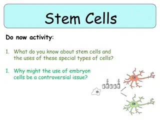
Understanding Hematopoiesis and White Blood Cells in Blood Formation Process

The process of hematopoiesis involves the lifelong production, multiplication, and specialization of different blood cells in the bone marrow, starting with the stem cells. This process includes the differentiation and proliferation of various progenitor cells, regulated by hematopoietic growth factors. Normal ranges of white blood cell counts and differential counts are crucial for assessing immune health. Neutrophils, a type of white blood cell, play a vital role in phagocytosis and immune response. Understanding chemotaxis and neutrophil extracellular traps sheds light on their role in fighting infections and inflammation.

Download Presentation

Please find below an Image/Link to download the presentation.
The content on the website is provided AS IS for your information and personal use only. It may not be sold, licensed, or shared on other websites without obtaining consent from the author. Download presentation by click this link. If you encounter any issues during the download, it is possible that the publisher has removed the file from their server.
E N D
Presentation Transcript
White Blood Cells White Blood Cells Dr. Mansour Aljabry Dr. Mansour Aljabry Assistant Professor & Assistant Professor & Consultant Hematologist Consultant Hematologist 1
Hematopoiesis Hematopoiesis is the process of life long production, multiplication & specialization of blood cells in the bone marrow. It begins with the most basic blood cell, the stem cell or pluripotent hematopoietic stem cell (PHSC). Proliferation Differentiation HSC (mother) 2
Lymphoid Progenitor Hematopoietic stem cell Granulocytic- Monocytic Progenitors Monocytes Myeloid Progenitor Erythroid Progenitor Megakaryocytic Erythroid Progenitors 3
Hematopoietic growth factors : Hormones that regulate the proliferation and differentiation of HCS. Can be administered clinically to stimulate the hematopoiesis : -G-CSF in severe neutropenia -EPO in Sever anemia 4
PAX-5 Lymphoid Progenitor T-bet Hematopoietic stem cell G-CSF SCF FLT3-L Granulocytic- Monocytic Progenitors Monocytes M-CSF EPO Myeloid Progenitor Erythroid Progenitor Megakaryocytic Erythroid Progenitors Thrombopoietin 5
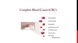
Normal ranges WBC count 4-11 x 109/L Absolute count (x109/L) Cells Differential count % Neutrophils 50-70 1.5-6.5 1.2-3.4 Lymphocyte 20-44 0.1-0.6 Monocytes 2-9 0-0.7 Bands 0-6 0-0.5 Eosinophils 0-5 0-0.2 Basophils 0-2 6
Neutrophil Phagocytic cell contain a nucleus divided into 2 5 lobes with pale granulated cytoplasm. stain a neutral pink (Basophils dark blue while eosinophil is bright red). They are short lived( 6 days) and highly motile. 8
Chemotaxis: Migration toward sites of infection or inflammation through detection of IL-8 ,TNF &C5a Neutrophil extracellular traps(NETs): are web-like structures of DNA and fiber that trap and kill microbes extracellularly Superoxide Hydrogen peroxide Lysozyme NADPH oxidase Myeloproxidase (MPO) 9
Neutrophil 10
Neutrophilia Marginal pool (tissues) Circulating pool (blood) 11
Causes of neutrophilia Benign Neoplastic Bacterial infection (Pyeogenic) Chronic Myeloid leukemia Polycythemia vera,Essential Thrombocythemia Inflammation and tissue necrosis Acute hemorrage or hemolysis Chronic Myelomonocytic Leukemia Steroid therapy (inhibits margination) Non hematological Neoplasms G-CSF treatment Rare inherited disorders Aspleni 12
Leukemoid reaction Leukocytosis(mainly neutrophilia with left shift) due to physiological response to stress or infection. Common with : sever or chronic infection ,sever hemolysis & metastatic cancers. Associated with: Toxic granulation ,Vaculation & Dohle bodies. 13
Left shift 14
Neutropenia Lower limit of normal is 1.5 10 /L Sever neutropenia : 0.5 10 /L Selective neutropenia Part of general pancytopenia Congenital : - Kostmann syndrome - Benign neutropenia , - Cyclical neutropenia Bone marrow failure Drug induced Chemotherapy Autoimmune (SLE) Acute Leukemia Viral infection (hepatitis ,HIV) Lymphoma Some Bacterial infections(Typhoid) Metastatic carcinoma 15
Lymphocytes Small WBC having spherical nucleus surrounded by a thin layer of non granular cytoplasm. Fundamental importance in the immune system (Innate &Adaptive) NK cells 5% B cells 15% T cells 80% 16
Lymphocyte (B or T) NK Lymphocyte Reactive looking Atypical looking 17
Lymphocytosis 1- Viral infection : Infectious mononucleosis ,cytomegalovirus ,rubella, hepatitis, adenoviruses, varicella . 2- Some bacterial infection: (Pertussis ,brucellosis ) 3- Other conditions: Allergic drug reactions, splenectomy, dermatitis ,hyperthyroidism metastatic carcinoma .) 4- Chronic lymphocytic leukemia (CLL) 5-Other lymphomas: Mantle cell lymphoma ,Hodgkin lymphoma 18
Lymphocytopenia Selective Lymphocytopenia Part of general pancytopenia Bacterial or fungal sepses Bone marrow failure Post operative state Chemotherapy or radiotherapy Post steroid therapy Acute Leukemia Immunodeficiency (congenital or acquired) Lymphoma Autoimmune disorders (SLE..) Metastatic carcinoma 19
Monocytes Large, phagocytic WBC, having a single well-defined nucleus and very fine granulation in the cytoplasm. Represents about 2-9% of WBC Matures into different types of macrophages at different anatomical locations (skin, spleen, liver ..) 20
Monocytes Monocytosis Monocytopenia Selective monocytopenia: 1- Chronic infection : (TB , Brucellosis..) Hairy cell leukemia 2-Autoimmune :SLE.. 3-Chronic neutropenia 4-Acute leukemia (AML M5) 5-Chronic Myelomonocytic leukemia 21
Eosinophil Granular leukocyte having a nucleus with two lobes and cytoplasm containing bright red coarse and round granules . Major functions : 1-Antiparasitic and bactericidal activity, 2-Participating in immediate allergic reactions, 3-Modulating inflammatory responses. 22
Eosinophilia Causes: C H I N A Connective tissue diseases Helminthic parasitic infections Idiopathic: Hypereosinophilic syndrome Neoplasia: eosinophilic leukaemia, lymphoma or MPN Allergy 23 23
Basophil Phagocytic leukocyte of characterized by numerous coarse bluish-black granules of variable size. Mature to tissue mast cells. Functions of Basophils - Major role in immediate allergic reaction (IgE) - Anticoagulant activity (Heparin) - Vasodilatation (Histamine) 24
Basophilia 1-Neoplasia (CML, mastocytosis, basophilic leukaemia, MPN) 2-Hypothyroidism 3-Some viral infections (Chickenpox) 4-Inflammatory conditions 5-Drugs (IL3, oestrogen) 6-Hyperlipidaemia 25 25
leukoerythroblastic picture Bone marrow response in which nucleated red blood cells and immature leukocytes are released in the blood Causes: Metastatic neoplasm Primary Myelofibrosis Severe hemolysis (Thalassemia major) Severe infection ( Miliary TB) 26