Overview of the Lymphatic System and Immune Response
The lymphatic system plays a crucial role in returning leaked fluids back to the blood, protecting the body from foreign materials, and supporting immune surveillance. It consists of lymphoid cells such as T and B lymphocytes, macrophages, dendritic cells, and reticular cells that work together to defend against antigens. The immune response involves T cells for cellular immunity and B cells for humoral immunity. Components of the lymphatic system include lymphatic vessels, capillaries, and lacteals, serving to maintain fluid balance and absorb fats from the intestines.

Download Presentation

Please find below an Image/Link to download the presentation.
The content on the website is provided AS IS for your information and personal use only. It may not be sold, licensed, or shared on other websites without obtaining consent from the author. Download presentation by click this link. If you encounter any issues during the download, it is possible that the publisher has removed the file from their server.
E N D
Presentation Transcript
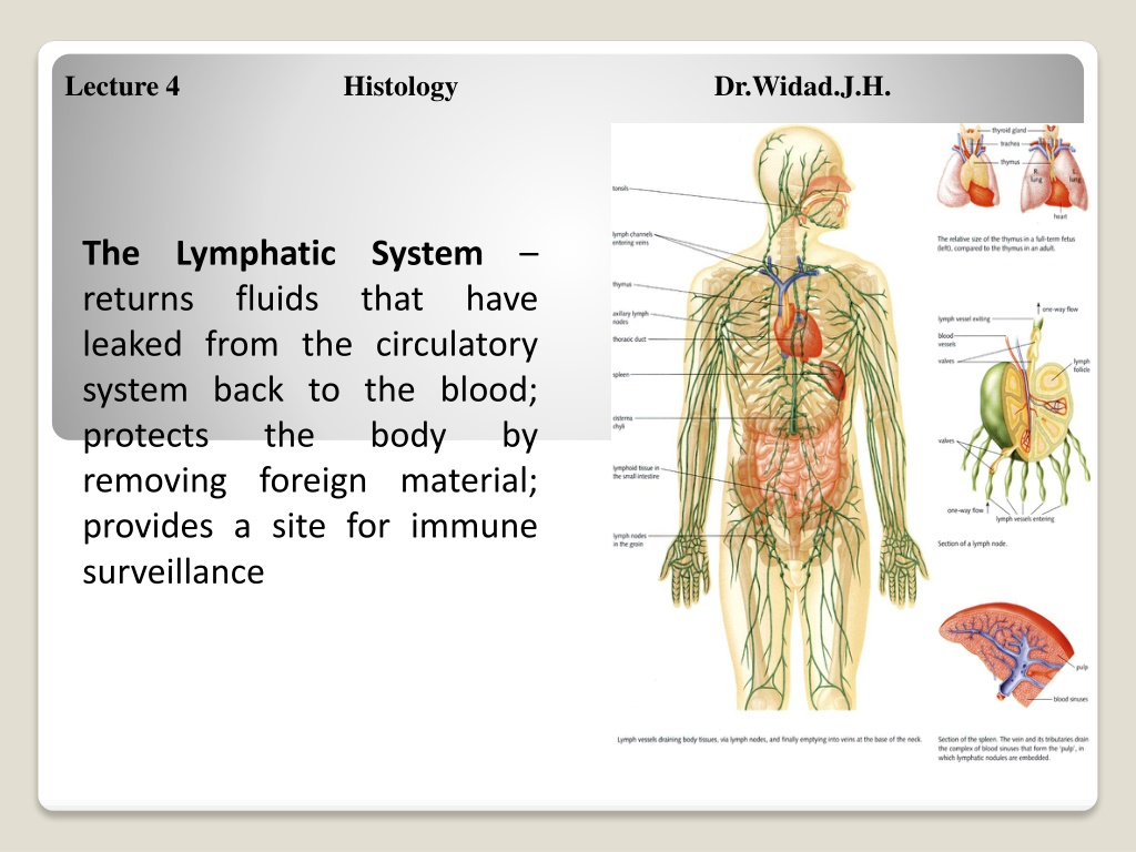
Lecture 4 Histology Dr.Widad.J.H. The Lymphatic System returns fluids that have leaked from the circulatory system back to the blood; protects the removing foreign material; provides a site for immune surveillance body by
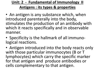
The Immune Response T cells = cellular immunity; function to amplify the inflammatory response B cells = humoral immunity T Cells and Cell Mediated Immunity - targets virus or parasite infected cells, cancer cells, and cells of foreign grafts 3 main types of cells: 1. Cytotoxic T (TC) cells: carry out cell mediated immunity, physically attack foreign cells 2. Helper T (TH) cells: activate B and TC cells 3. Suppressor T (TS) cells: moderate the immune response by inhibiting TC and B cells
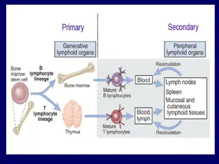
Lymphoid cells Lymphocytes Arise in the red bone marrow Protect the body against antigens Two types T lymphocytes (T cells) oMature in the thymus oDirectly attack and destroy foreign cells B lymphocytes (B cells) oMature in the bone marrow oProduce manufacture antibodies plasma cells that
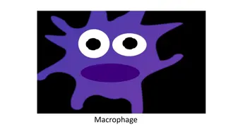
Macrophages substances and activate the T cell response Dendritic cells initiate the immune response Reticular cells produce the reticular fibers that form the soft skeletal structure of lymphoid organs phagocytize foreign
Components of the Lymphatic System Lymphatic vessels return to the blood any fluids that have escaped from the circulation Distribution of lymphatic vessels Lymphatic vessels travel alongside blood vessels Lymphatic vessels are absent from bones, teeth, bone marrow, and the central nervous system Lymphatic capillaries microscopic blind-ended tubes that are interwoven between the tissue cells and the blood capillaries Lacteals specialized lymphatic capillaries of the intestinal mucosa Fatty lymph (chyme) containing fats and fat-soluble substances is absorbed in the lacteals
Lymph transport The lymphatic circulation is a low-pressure system The lymphatic system lacks a pumping organ; must utilize the valves, respiratory pumps and muscular pumps to promote lymph flow toward the heart The movement of surrounding tissues is also important in propelling lymph through the lymphatics The lymphatic capillaries converge into larger vessels Lymph flows from the lymphatic capillaries to lymphatic collecting vessels to lymphatic trunks to the right lymphatic duct/thoracic duct
The right lymphatic duct drains lymph from the right upper arm and the right side of the head and thorax The thoracic duct receives lymph from the rest of the body
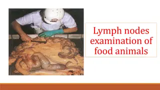
Lymph nodes Only the lymph nodes filter lymph Cluster along the lymphatic vessels of the body Lymph is filtered through the lymph nodes before it is returned to the bloodstream Lymph nodes are embedded in connective tissue Large clusters of lymph nodes appear near the body surface in the inguinal, axillary, and cervical regions. Functions of lymph nodes Filters lymph Assist in activating the immune system
Anatomy of a lymph node Most are kidney-shaped Surrounded by a fibrous capsule Strands of connective tissue (trabeculae) divide the node into compartments Two histologically distinct regions Cortex oContains densely packed follicles with many germinal centers oDeeper portion of the cortex primarily houses T cells Medulla oContain both types of lymphocytes
Circulation in the lymph nodes Afferent lymphatic vessels lymph enters here Once inside the nodes, the lymph moves through a series of sinuses and then exits at the hilus Efferent lymphatic vessels lymph exits out the nodes
Spleen The largest lymphoid organ Site of lymphocyte proliferation and immune surveillance and response Functions of the spleen Cleanses the blood by removing old RBCs and platelets, as well as debris from the blood. Stores the breakdown products of RBCs Site of erythrocyte production in the fetus
Anatomy of the spleen Surrounded by a fibrous capsule Contains both T cells, B cells, RBCs and macrophages Divided histologically into two regions Red pulp rich in lymphocytes and reticular fibers oRBC disposal and recycling White pulp rich in macrophages and reticular fibers oImmune functions
Thymus the site of T cell maturation Most active in younger children; atrophies with age Does not contain reticular fiber Lack B cells, therefore no germinal cells centers are present in the thymus
. Thymus 1. Location - situated above the heart where the great vessels connect. 2. Importance - during early life when the cellular mediated component of the immune system develops. Undergoes atrophy in later life, at which time it loses its functional significance. 3. The thymus consists of multiple lobes each containing characteristic cortical and medullary structure; however, these are not lymphatic nodules (i.e., not a spherical structure that is distinct from surrounding cells). A connective tissue capsule surrounds the thymus.
a. Epithelial tissues of embryos pharynx are internalized during development and move to site of thymus dorsal to b. These epithelial tissues are invaded by lymphoblasts (immature T-lymphocytes) that originate from stem cells in the bone marrow. c. The invading cells organize themselves into the cortical and medullary portions of lobules.
* Cortical area consists of dense population of so-called thymocytes, that are T-lymphocytes, and scattered epithelial reticular cells that have multiple processes and partially compartmentalize the thymocytes. * These cells surround a central zone of loose lymphatic tissue called the medullary zone that consists of fewer thymocytes and more epithelial reticular cells. * The cortical and medullary zones of lobules are all continuous with each other.
d. Other cell types found in the thymus are: * macrophages * plasma cells * mast cells 5. Cortical layer of thymus a. Site of lymphocyte production - divisions of lymphoblast cells. b. Thus, there is considerable mitotic activity of lymphoblasts c. Epithelial reticular cells are less numerous in this area and have thin and long processes that evelope groups of developing thymocytes. No reticular fibers are present
. Medullary zone a. Contains mostly epithelial-reticular cells and fewer T- lymphoblasts and lymphocytes than the cortex. b. Also contains specialized structures known as Hassall's corpuscles - function unknown * consist of a central, eosinophilic, hyaline core surrounded by concentric layers of epithelial reticular cells containing lots of keratin. *sometimes they are calcified. *these structures are characteristic of thymus.
1. Palatine tonsils a. On left and right in rear area of oral cavity. b. Dense lymphoid tissue that forms a band of lymphatic nodules that lie just below a non-keratinized, stratified, squamous epithelium lining the oral cavity in this region. c. Overlying epithelium forms invaginations called multiple crypts that penetrate into the band of nodules. d. These crypts act as collecting places for cellular debris and bacteria as well as some living lymphocytes that have migrated into the crypts. e. The band of lymph nodules is separated from underlying tissues by a partial capsule of dense connective tissue.
Tonsils the simplest lymphoid organs; named according to their location Palatine tonsils Lingual tonsils Pharyngeal tonsil Tubal tonsils
2. Pharyngeal tonsils a. Diffuse lymphoid tissue containing nodules, but no crypts. b. Mostly lie beneath a typical pseudostratified ciliated columnar respiratory epithelium in rear roof of pharynx. Some areas of the covering epithelium may be stratified squamous. c. A thin partial capsule of dense connective tissue separates the lymphoid tissue from underlying tissue.
2. Pharyngeal tonsils a. Diffuse lymphoid tissue containing nodules, but no crypts. b. Mostly lie beneath a typical pseudostratified ciliated columnar respiratory epithelium in rear roof of pharynx. Some areas of the covering epithelium may be stratified squamous. c. A thin partial capsule of dense connective tissue separates the lymphoid tissue from underlying tissue.
3. Lingual tonsils a. Situated in the root of tongue. b. Each lingual tonsil consists of numerous. lymphoid nodules surrounding a single crypt c. The crypt is lined by a non-keratinized, stratified, squamous epithelium. d. A thin partial capsule of dense connective tissue separates the lymphoid tissue from underlying tissue
Aggregates of lymphoid follicles Location of these follicles make them ideal because they are able to: Destroy bacteria and prevent pathogens from slipping through the intestinal wall Generate many memory lymphocytes for long- term immunity Examples Peyer s patches found in the distal portion of the small intestine Appendix of the cecum (the first part of the large intestine)