Comprehensive Imaging of Wrist by Dr. Amita Harsule
This collection of images showcases various radiographs and ultrasound scans of the wrist, focusing on different angles and structures. Detailed descriptions accompany the images, highlighting specific findings such as disruptions in joint arcs and abnormal step-offs. The comprehensive imaging provides valuable insights into wrist conditions and pathologies that may be present, aiding in diagnosis and treatment planning.
Download Presentation

Please find below an Image/Link to download the presentation.
The content on the website is provided AS IS for your information and personal use only. It may not be sold, licensed, or shared on other websites without obtaining consent from the author. Download presentation by click this link. If you encounter any issues during the download, it is possible that the publisher has removed the file from their server.
E N D
Presentation Transcript
Imaging of Wrist DR AMITA HARSULE
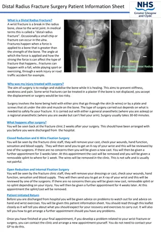
Basic Radiograph ROUTINE PA Lateral Scaphoid series when there is tenderness in the anatomical snuff box.
PA ulnar deviation
note the disruption of arc I at the lunotriquetral joint.
A disruption of the second carpal arc at the scapholunate joint and the lunotriquetral joint is seen on the left.
There is an abnormal step off at the capitohamate joint.
Volar aspect of the wrist
Ligamentous Anatomy Classified as either extrinsic or intrinsic. The extrinsic ligaments link the carpal bones to the radius and ulna. The intrinsic or intercarpal ligaments connect the individual carpal bones.
Normal TFCC. Triangular fibrocartilage proper (*). Foveal (arrow) and styloid (dashed arrow) insertions. Meniscus homologue (arrow head). Normal lunotriquetral (LTL) and scapholunate (SLL) ligaments are also seen.
Tendon pathology Tendon pathology is often encountered in the wrist and hand, which may be the result of overuse, trauma or inflammation.Patients typically present with focal pain and swelling. The term "tendinitis" is an inaccurate term A better term is tendinosis, which describesa degenerative process with "microtears", healing and vascularin-growth. The term "tendinopathy" encompasses both degenerativeand inflammatory conditions. Tenosynovitis refers to inflammatorychange within the tendon sheath, with or without tendon morphologicalchanges.
flexor tenosynovitis
Carpal Tunnel $ Causes include flexor tenosynovitis, trauma, systemic inflammatory disease, pregnancy, acromegaby, hypothyroidism
Space-occupying lesions within the carpal tunnel such as ganglia, tenosynovitis, and anatomic variants are well seen with MR imaging MR imaging is therefore indicated only in patients with equivocal clinical findings, a suspected mass
Compressive ulnar neuropathy. (A) At the level of the canal of Guyon s. The Guyon canal is a triangular space lateral to the pisiform bone and medial to the hamate that contains the ulnar nerve and ulnar artery. (B) The ulnar nerve (arrow) has high signal intensity and it is thickened. There is a ganglion cyst (*) compressing the ulnar nerve against the pisiform. (C)
GANGUON CYSTS Seventy percent of all wrist ganglia occur dorsally adjacent to the SLL
1. ganglion cyst that arises from the dorsal aspect of the SLL 2. volar ganglion cyst adjacent to the radioscaphoid joint
schwannoma of radial nerve
