Understanding Digital Radiography in Modern Healthcare
Digital radiography revolutionizes imaging by capturing, displaying, and storing radiographic images digitally, offering benefits such as dose reduction, image manipulation, and electronic transfer. While there are advantages like environmentally friendliness, it also comes with challenges like high initial costs and storage requirements. Different methods like direct and indirect sensors, as well as Storage Phosphor Imaging, play crucial roles in this advanced imaging technique.
Download Presentation
Please find below an Image/Link to download the presentation.
The content on the website is provided AS IS for your information and personal use only. It may not be sold, licensed, or shared on other websites without obtaining consent from the author. Download presentation by click this link. If you encounter any issues during the download, it is possible that the publisher has removed the file from their server.

Presentation Transcript
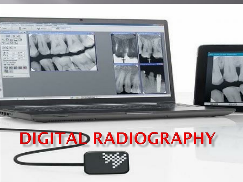
Digital radiography is a term used to describe radiography when the images are taken in a digital form and are capable of being displayed in the computer Digital sensors are used
Digital radiography refers to a method of capturing a radiographic image using a sensor, breaking it into electronic pieces and presenting and storing the image using a computer.
Dose reduction upto 50 -90% compared to conventional Image can be manipulated Contrast of the image can be enhanced Measurements can be done Images are electronically transferred to other healthcare providers It can stored for future use and study purpose Environment friendly avoids chemicals used in processing and lead foil
Relatively high initial cost Rigid sensor dimensions Large disc space is needed to store the data
Direct - sensor is placed in the patients mouth and exposed to radiation. From the sensor the image is transmitted to computer monitor within seconds Indirect - an existing X-ray film is digitized using a CCD camera, which scans the image, digitizes or converts the image and displays in computer.
Storage Phosphor Imaging A reusable imaging plate coated with phosphors is used. The storage phosphor imaging records diagnostic data on the plates following exposure to the X-ray source and high speed scanner to convert the information to electronic files --- displayed on the computer screen
X-ray generator Direct digital sensors Charge coupled device (CCD) Complementary metal oxide semiconductors (CMOS) Photostimulable phosphor plates (PSP) Thin film transistors (TFT) Analog to digital converter (ADC) Computer Monitor or printer for Image display
Matrix the image to be taken is represented as many rows and columns Pixel picture element. Each pixel is assigned a value, related to signal intensity. The value stored in a binary form (0,1) Pixel having high value represents dark gray shade, whereas low value represents white shade. High dose high value dark gray shade
Image acquisition Image processing Image display Image Archiving Image retrieving
Digital radiography Digital radiography Computed Radigraphy CCD Indirect flat panel detector Direct Flat panel PSP
First adapted in dentistry in 1987 It is an integrated circuit made of crystalline silicon Forms images from a visible light Uses the same radiographic machine as the conventional Uses sensors instead of radiographic film to capture
CCD uses a thin wafer of silicon as the basis for image recording. The silicon crystals are formed in a picture element (pixel) matrix. When exposed to radiation, the covalent bonds between silicon atoms are broken, producing electron hole pairs. Electrons are the attracted toward the most positive potential in the device, where they create charge packets Each packet corresponds to one pixel
The image is read by transferring each row of pixel charges from one pixel to the next in a bucket brigade fashion
3 components component Function Scintillator Convert X-ray into visible light Fiberoptic Transmit visible light to chip and stop X-rays Main CCD component Convert light signal to electric voltage
Fiberoptic scintillator CCD component e- e- e- e- ADC X-rays e- e- e- e- e- Light waves computer
After passing through the patient the x- ray first strikes on the scintillator It converts the X-rays to visible light Visible light passes through the fiberoptic This transmits light to CCD CCD consists of multiple small elements called pixels (picture element)
Visible light is absorbed in individual pixel With the help of small transistor element made of silicon, the electrons are released The electrons are deposited in the electron well itself Once CCD is exposed, charge is read out in Bucket brigade fashion i.e. The electronic charge is shifted from pixel by pixel at the boundaries and then collected These electrons are converted to electric voltage signal
Analog signal is sent to the analog digital converter (ADC) ADC converts analog image to numeric values This is then passed on to computer and converted to a visible image on a monitor Electrons stored will be proportional to the number of X-rays photon strike the element