Anatomy of Hand and Wrist: Structures and Functions Overview
This lecture covers the anatomy of the hand and wrist, focusing on the deep fascia, flexor and extensor retinacula, and the insertion of tendons. It explores the structures passing superficial and deep to the flexor retinaculum and the small muscles of the hand. Important details about the carpal tunnel, including causes and manifestations of carpal tunnel syndrome, are also discussed.

Download Presentation

Please find below an Image/Link to download the presentation.
The content on the website is provided AS IS for your information and personal use only. It may not be sold, licensed, or shared on other websites without obtaining consent from the author. Download presentation by click this link. If you encounter any issues during the download, it is possible that the publisher has removed the file from their server.
E N D
Presentation Transcript
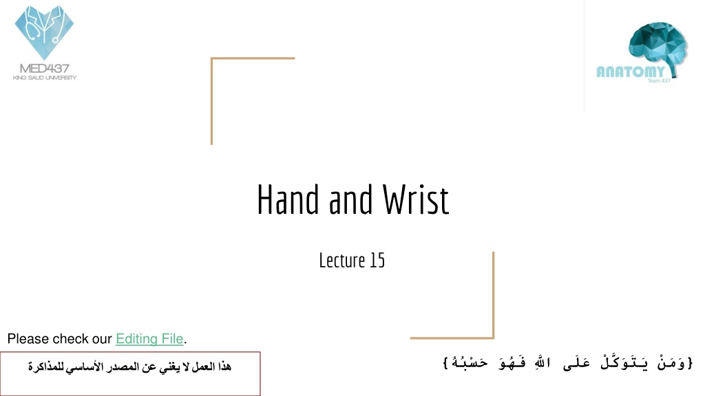
Hand and Wrist Lecture 15 Please check our Editing File. { }
Objectives Describe the anatomy of the deep fascia of the wrist & hand (flexor & extensor retinacula & palmar aponeurosis). List the structures passing superficial & deep to flexor retinaculum. Describe the anatomy of the insertion of long flexor & extensor tendons. Describe the anatomy of the small muscles of the hand (origin, insertion action & nerve supply) Text in BLUEwas found only in the boys slides Text in PINKwas found only in the girls slides Text in RED is considered important Text in GREY is considered extra notes
Retinacula Retinaculum=single | Retinacula=plural Flexor & Extensor Retinaculum: Bands of Deep (thickening) Fascia at the Wrist Hold the long flexor and extensor tendons at the wrist in position. Function Medially: Both retinacula attached to Pisiform & Hook of Hamate. Laterally: - Flexor Retinaculum attached to Tubercle of Scaphoid & Trapezium. - Extensor Retinaculum attached to Distal end of Radius. Attachment
Structure superficial to flexor Retinaculum From Medial to Lateral 1. Tendon of Flexor carpi ulnaris. 2. Ulnar nerve. 3. Ulnar artery. 4. Palmar cutaneous branch of ulnar nerve. 5. Palmaris longus tendon. 6. Palmar cutaneous branch of median nerve. Do NOT enter the carpal tunnel
*Paresthesia refers to a burning or prickling sensation that is usually felt in the hands, arms, legs, or feet, but can also occur in other parts of the body. Carpal Tunnel from Concave anterior surface of the Carpus covered by Flexor Retinaculum. Form From Medial to Lateral Tendons of flexor digitorum superficialis & profundus Median nerve Flexor Pollicis Longus Flexor carpi radialis Content Causes : Compression of the median nerve within the carpal tunnel. Manifestations: 1. Burning pain (pins and needles ) in the lateral three and half fingers. 2. No paresthesia* over the thenar eminence Because the palmar cutaneous of median nerve does not go under the tunnel 3. Weakness or atrophy of the thenar muscles (Ape Hand). 4. Inability to Oppose the thumb. Carpal tunnel syndrome
Palmar Aponeurosis - The Thickened deep fascia of the Palm. - it is Triangular in shape , occupies the central area of the palm. Attached to the distal border of flexor retinaculum and receives the insertion of palmaris longus tendon. Apex Divides at the bases of the fingers into four slips that pass into the fingers Base 1. Firmly attached to the overlying skin and improves the grip. 2. Protects the underlying tendons, vessels & nerves. 3. Gives origin to palmaris brevis muscle. Functions
Small muscles of the hand Palmaris Brevis Flexor retinaculum (FR) & Palmar aponeurosis (PA) Origin Skin of the palm. Insertion Ulnar nerve (superficial branch) Nerve supply Corrugation of skin to improve grip. Action
Short muscles of hand (thumb & little finger) + Movement of thumb Team436 Can be remembered using the mnemonic, "A OF A OF A" for: (thenar muscles) Abductor pollicis brevis Opponens pollicis Flexor pollicis brevis Adductor pollicis (Hypothenar muscles) Opponens digiti minimi Flexor digiti minimi Abductor digiti minimi
Small muscles of the hand Hypothenar Eminence (3 muscles) Abductor Digiti minimi Flexor Digiti Minimi Opponens Digiti Minimi Pisiform Flexor retinaculum Flexor retinaculum Origin Base of proximal phalanx Base of proximal phalanx Palmer surface of 5th metacarpal Insertion Deep branch of ulnar nerve Deep branch of ulnar nerve A Deep branch of ulnar nerve Nerve supply Abduction of little finger Flexion of little finger Pulls the 5th metacarpal forward Action
Small muscles of the hand Thenar Eminence (3 muscles) Abductor Pollicis Brevis Flexor Pollicis Brevis Opponens Pollicis Flexor retinaculum, Scaphoid and trapezium Flexor retinaculum Flexor retinaculum Origin Base of proximal phalanx Base of proximal phalanx Lateral part of 1st metacarpal Insertion Supplied by median nerve Supplied by median nerve Supplied by median nerve Nerve supply Abduction of thumb Flexion of thumb Opposition of thumb Action
Small muscles of the hand Not in the Thenar Eminence
Small muscles of the hand Writing Position
Insertion of: Each tendon: - Divides into two halves & pass around the Profundus Tendon. - The two halves Meet on the posterior aspect of Profundus tendon (partial decussation of fibers). - Reunion of the two halves. - Further Division into two slips attached to the Borders of Middle Phalanx. Flexor dig superficialis Inserted into the Base of the Distal Phalanx. Flexor dig Profundus Green -> superficialis |Grey -> Profundus
Fibrous flexor (digital) sheath Fibrous Flexor is strong fibrous sheath which covers the anterior surface of the fingers and attached to the sides of the phalanges . Its proximal end is opened. Its distal end is closed. The sheath with the anterior surface of the phalanges and interphalangeal joints form osteofibrous blind tunnel for the long Flexor tendons of fingers.
Synovial flexor sheath A- Common synovial sheath ( Ulnar bursa ) Contains tendons of Flexor digitorum superficialis and profundus. The medial part of the sheath extends distantly ( without interruption) on the tendons of the little finger. The lateral part of the sheath stops on the middle of the palm. (doesn't cover the 3 middle fingers) The distal ends of the long Flexor tendons to - index, middle and ring - fingers acquire digital synovial sheaths. B- Flexor Pollicis Longus Tendon has its own synovial sheath ( Radial bursa) They allow the long tendons to move smoothly with a minimum of friction beneath the Flexor retinaculum and the fibrous Flexor sheaths Functions
Extensor Expansion - Formed from the expansion of the tendons of extensor digitorum at the PIJ (P = proximal - I = interphalangeal - J = joint) - The tendon splits into three parts: One Central: inserted into the base of Middle phalanx. Two laterals: inserted into the base of the Distal phalanx. - The Expansion Receives the insertions of: Corresponding Interosseous muscle (on each side). Lumbrical muscle (on the lateral side).
Action of Lumbricals & Interossei Writing position
MCQs (4) Which one of the following is a symptom of Carpal Tunnel Syndrome _______ : A- burning pain B- weakness C- both A and B (1) Which one of the following is Hypothenar Eminence ______ : A- abductor digiti minimi B- opponens digiti minimi C- both A & B (5) Retinacula bands of ______ Fascia at the Wrist : A- deep B- superficial C- both A and B SAQ (1) List the attachment Retinacula? (2) Each tendon of flexor digiti superficialis divides into ______ around the profundus tendon : A- 2 B- 3 C- 4 (3) In Fibrous Flexor (Digital) Sheath the proximal end is ______ and the distal end is ______ : A- closed, opened B- opened, closed C- opened, opened (2) A boy injured his median nerve and as a result there was a wasting in the thenar muscles. List the muscles affected and the action of each one? (3) Lost the Structure superficial to flexor Retinaculum from medial to lateral?
Answers SAQ: (1) Medially: Both retinacula attached to Pisiform & Hook of Hamate. Laterally: - Flexor Retinaculum attached to Tubercle of Scaphoid & Trapezium. - Extensor Retinaculum attached to Distal end of Radius. (MCQs) 1-B 2-A 3-B 4-C 5-A (2) 1) abductor pollicis brevis (abduction) 2) flexor pollicis brevis (flexion) 3) opponens pollicis (opposition) (3) 1. Tendon of Flexor carpi ulnaris. 2. Ulnar nerve. 3. Ulnar artery. 4. Palmar cutaneous branch of ulnar nerve. 5. Palmaris longus tendon. 6. Palmar cutaneous branch of median nerve.
Team Members Lamia Abdullah Alkuwaiz (Team Leader) Rawan Mohammad Alharbi Abeer Alabduljabbar Afnan Abdulaziz Almustafa Ahad Algrain Alanoud Almansour Albandari Alshaye AlFhadah abdullah alsaleem Arwa Alzahrani Dana Abdulaziz Alrasheed Dimah Khalid Alaraifi Ghada Alhaidari Ghada Almuhanna Ghaida Alsanad Hadeel Khalid Awartani Haifa Alessa Khulood Alwehabi Layan Hassan Alwatban Lojain Azizalrahman Lujain Tariq AlZaid Faisal Fahad Alsaif (Team Leader) Abdulaziz Al dukhayel Maha Barakah Majd Khalid AlBarrak Norah Alharbi Nouf Alotaibi Noura Mohammed Alothaim Rahaf Turki Alshammari Reham Alhalabi Rinad Musaed Alghoraiby Sara Alsultan Shahad Alzahrani Wafa Alotaibi Wejdan Fahad Albadrani Wjdan AlShamry Abdulmajeed Alwardi Abdulrahman Alageel Rayyan Almousa Sultan Alfuhaid Ali Alammari Fahad alshughaithry Fayez Ghiyath Aldarsouni Mohammed Alquwayfili Fahad Alfaiz Akram Alfandi Saad Aloqile Saleh Almoaiqel Abdulaziz Alabdulkareem Abdullah Almeaither Yazeed Aldossari Muath Alhumood Abdulrahman Almotairi Abduljabbar Al-yamani Sultan Al-nasser Majed Aljohani Zeyad Al-khenaizan Mohammed Nouri Abdulaziz Al-drgam Fahad Aldhowaihy Omar alyabis Abdulelah Aldossari Abdulrahman Alduhayyim Hamdan Aldossari Abdullah Alqarni Mohammed Alomar Abdulrahman Aldawood Saud Alghufaily Hassan Aloraini Khalid Almutairi


")


 sheath")