Understanding the General Characteristics of Gymnosperms
Gymnosperms, originating in the Paleozoic era, were dominant in the Jurassic and Cretaceous periods. With naked seeds and xerophytic traits, these plants exhibit unique features such as heterospory, symbiotic root associations, and different leaf types. Examples include Cycus, Pinus, and Gnetum. Their anatomical structure includes thick cuticles and differentiated mesophyll tissues. Explore the distinct traits and evolutionary history of gymnosperms in this detailed overview.
Download Presentation

Please find below an Image/Link to download the presentation.
The content on the website is provided AS IS for your information and personal use only. It may not be sold, licensed, or shared on other websites without obtaining consent from the author. Download presentation by click this link. If you encounter any issues during the download, it is possible that the publisher has removed the file from their server.
E N D
Presentation Transcript
Gymnosperms Class Teacher Dr. Hannan Mukhtar Assistant Professor Department of Botany Lahore College for Women University, Lahore Pakistan. Course Description Course Title: Course code: Credit hours: DIVERSITY OF PLANTS Min/Bot-102 4 (3+1) Class Course : Major: Minor course: BS I, 2ndSemester Zoology Botany
General characteristics of Gymnosperms Gymnosperms were originated in Paleozoic era (541-252 million years ago). They were the dominant plants of the Jurassic and Cretaceous periods of the Mezozoic era. Most ancient seed bearing vascular plants containing 83 genera and 1080 species. Many primitive gymnosperms are extinct. Examples of living Gymnosperms Cycus, Pinus, Gnetum, Ephedra, Welwitshi, Texus, Cedrus, Abies and Araucaria Morphological Characters Gymnosperms (gymnos- naked and sperm- seed) are naked seeded plants. Mostly are ever green woody, perennials with shrubby or tree habit They show xerophytic chracteristics. Mostly are very large in size. Ovules are present on scales which are arranged to form a cone but ovules are exposed.
General characteristics of Gymnosperms The plant body is sporophyte (diploid) differentiated into roots stems and leaves. Gymnosperms are hetrosporous, produce two types of spores i.e. microspores and megaspore Two types of sporangia are present microsporangia and megasporangia (pollen sac) which are produced on specialized leaves i.e. sporophylls. Two types of sporophylls are present i.e. microsporophyll (stamen) contaning microsporangia and megasporophyll (carpel) containing mega sporangium (ovule). The roots show symbiotic association with fungi and cyanobacteria. Fungi form mychorrhizal association with the roots of Pinus. Mychorrhiza helps in absorption of minerals. Algae ( Anabena and Nostoc) inhibits in the coralloid roots of Cycas , Which helps in nitrogen fixation. The stem is usually erect branched and woody, unbranched in cycus underground in Zamia
General characteristics of Gymnosperms Presence of leaf scar is the characteristic feature of gymnosperms. Leaves are usually dimorphic ( two types of leaves in same plants) That are foliage and scale leaves. Foliage leaves are green simple, simple or needle shaped or pinnately compound leaves. Scale leaves are minute and decidous. Cycus show circinate vernation Presence of cercinate vernation in Cycus is the strong evidence of Pteridophytic origin of gymnosperms . Cycus is a link between Pteridophytes and Gymnosperms. Anatomical characters The leaves of gymnosperms have very thick cuticle. Mesophyll is differentiated into spongy and palisade tissue.
https://www.google.com/search?q=cercinate+vernation+in +zimia&tbm https://www.google.com/search?q=foliage+and+scally++leaves+ of+pinus&tbm
Sunken stomata are present Have well developed vascular system consisting upon xylem and phloem Vascular bundles are open and collateral. Xylem Consists of tracheid and parenchyma Vessels are absent Vessels are present in wood of Gnetum, is a strong evidence for the gymnospermic origin of Angiosperms. Gnetum is a connecting link between Angiosperms and gymnosperms. Phloem Consists of sieve tubes and phloem parenchyma. Companion cells are absent. Stem shows secondary growth Wood may be manoxylic or pycnoxylic.
https://www.google.com/search?q=transfussion+tissue+in+gy mnosperms
Tenniniferous cells present in cortex. Roots are diarch (two) to polyarch (many) vascular bundles. Reproduction in Gymnosperms Hetrospory Microsporangia are produced on Microsporophyll. Megasporangia are produced on Megasporophyll. Sporophylls aggregates to form cone or stobili. Cones/ strobili are monosporangeate. Male cone, Microspores and Microsporophyll Male cones are short live. Microsporangia are formed on the abaxial side of microsporophyll. Microsporangium development is eusporangiate. https://www.google.com/search?q=cycas+male+and+female+cone+dia gram
Female cone, megasporophyll and mega sporangium Female cone is formed by aggregation of megasporophylls. Megasporophylls may be foliar as in Cycas or cauline (woody) as in Pinus. Megasporangium is ovule Ovular integument in gymnosperms is three layered. Pollination and fertilization Pollination by wind (Anemophily) Pollen deposit in wet pollen chamber Pollination siphonogamous (by pollen tube) Male gametes non motile except in Ginko and Cycas Number of archegonia in female gametophyte may vary. There are several archegonia in Cycas only one in pinus. Archegonium has ovum and neck canal cells (NCC) Absent in Gnetum.
https://www.google.com/search?q=microsporangium+with+microsporehttps://www.google.com/search?q=microsporangium+with+microspore +cycas
Embryo development is meroblastic (embryo develops from some part of zygote) Endosperm is present in seeds of gymnosperms. Endosperm is haploid (part of female gametophyte) Double fertilization absent Polyembryony is very common Integument- seed coat Seed are light weight and winged. Alternation of generation is present Diploid sporophytic generation is dominant phase. Haploid gametophytic generation is highly reduced. Gametophyte is dependant upon sporophyte. https://www.easybiologyclass.com/general-characters-of-gymnosperms- lecture-notes-with-ppt/
Life cycle of Gymnosperms https://www.google.com/search?q=general+characters+of+gymnosp erms
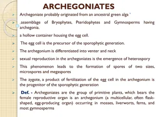
PINUS Kingdom Plantae Subkingdom Tracheobionta Superdivision Spermatophyta Division Coniferophyta Class Pinopsida Order Pinales Family Pinaceae Genus Pinus L.
External Morphology of Pinus Pinus is a large, perennial, evergreen plant. Branches grow spirally and thus the plant gives the appearance of a conical or pyramidal structure. Sporophytic plant body is differentiated into roots, stem and acicular (needle-like) leaves (Fig. 26). A tap root with few root hair is present but it disappears soon. Later on many lateral roots develop, which help in absorption and fixation The ultimate branches of these roots are covered by a covering of fungal hyphae called ectotrophic mycorrhiza. The stem is cylindrical and erect, and remains covered with bark. Branching is monopodial. Two types of branches are present: long shoots and dwarf shoots. These are also known as branches of unlimited and limited growth, respectively
Long shoots contain apical bud and grow indefinitely. Many scaly leaves are present on the long shoot. Dwarf shoots are devoid of any apical bud and thus are limited in their growth. They arise on the long shoot in the axil of scaly leaves. A dwarf shoot (Fig. 27) has two scaly leaves called prophylls, followed by 5-13 cataphylls arranged in 2/5 phyllotaxy, and 1-5 needles. The leaves are of two types, i.e., foliage and scaly. Scaly leaves are thin, brown-coloured and scale like and develop only on long as well as dwarf shoots. Foliage leaves are present at the apex of the dwarf shoots only. Foliage leaves are large, needle-like, and vary in number from 1 to 5 in different species. A spur (Fig. 28) is called unifoliar if only one leaf is present at the apex of the dwarf shoot, bifoliar if two leaves are present, trifoliar if three leaves are present, and so on.
Foliage leaves are large, needle-like, and vary in number from 1 to 5 in different species. 15. A spur is called unifoliar if only one leaf is present at the apex of the dwarf shoot, bifoliar if two leaves are present, trifoliar if three leaves are present, and so on. Pinus Species Pinus monophylla P. sylvestris P. gerardiana P. quadrifolia P. wallichiana-pentafoliar
Anatomical study T.S. Root Showing Secondary Growth: On the outer side are present a few layers of cork, formed by the meristematic activity of the cork cambium. Cork cambium cuts secondary cortex towards inner side. Many resin canals and stone cells are present in the secondary cortex, the cells of which are separated with the intercellular spaces. Below the phloem patches develop cambium, which cuts secondary phloem towards outer side and secondary xylem towards inner side. Crushed primary phloem is present outside the secondary phloem (Fig. 30). Many uniseriate medullary rays are present in the secondary xylem. Primary xylem is the same as in young roots, i.e., each group is bifurcated (Y-shaped) and a resin canal is present in between the bifurcation.
T.S. Long Shoot : Secondary growth, similar to that of a dicotyledonous stem, is present in the old stem of Pinus. Cork cambium cuts cork towards outer side and a few layers of secondary cortex towards inner side. Many tannin-filled cells and resin canals are distributed in the primary cortex. Cambium cuts secondary phloem towards outer side and secondary xylem towards inner side (Fig. 32). Primary phloem is crushed and pushed towards outer side by the secondary phloem. In the secondary xylem, annual rings of thin-walled spring wood (formed in spring season) and thick- walled autumn wood (formed in autumn season) are present alternately. Such a compact wood is called pycnoxylic (Age of the plant can be calculated by counting the number of these annual rings).
Below the secondary xylem are present a few groups of endarch primary xylem. Some of the medullary rays connect the pith with the cortex and called primary medullary rays while the others run in between secondary xylem and secondary phloem and called secondary medullary rays. Central part of the stem is filled with the parenchymatous pith. Resin canals are present in cortex, secondary xylem, primary xylem and rarely in the pith
T.S. Needle (Foliage Leaf): It is circular in outline in Pinus monophylla, semicircular in P. sylvestris and triangular (Fig. 38) in P. longifolia, P. roxburghii, etc. Outermost layer is epidermis, which consists of thick-walled cells. It is covered by a very strong cuticle. Many sunken stomata are present on the epidermis (Fig. 38). Each stoma opens internally into a substomatal cavity and externally into a respiratory cavity or vestibule. Below the epidermis are present a few layers of thick- walled sclerenchymatous hypodermis. It is well-developed at ridges. In between the hypodermis and endodermis is present the mesophyll tissue.
Cells of the mesophyll are polygonal and filled with chloroplasts. Many peg-like infoldings of cellulose also arise from the inner side of the wall of mesophyll cells. Few resin canals are present in the mesophyll, adjoining the hypodermis. Their number is variable but generally they are two in number. Endodermis is single-layered with barrel-shaped cells and clear casparian strips. Pericycle is multilayered and consists of mainly parenchymatous cells and some sclerenchymatous cells forming T-shaped girder, which separates two vascular bundles (Fig. 38). Transfusion tissue consists of tracheidial cells. Two conjoint and collateral vascular bundles are present in the centre. These are closed but cambium may also present in the sections passing through the base of the needle. Xylem lies towards the angular side and the phloem towards the convex side of the needle.
Reproductive Structures of Pinus: Plant body is sporophytic. Pinus is monoecious, and male and female flowers are present in the form of cones or strobili on the separate branches of the same plant. Many male cones are present together in the form of clusters, each of which consists of many microsporophylls. The female cones consist of megasporophylls. The male cones on the plant develop much earlier than the female cones. Male Cone: Separate a male cone from the cluster, study its structure, cut its longitudinal section, study the structure of a single microsporophyll, and also prepare a slide of pollen grains and study. The male cones develop in clusters (Fig. 39) in the axil of scaly leaves on long shoot. They replace the dwarf shoots of the long shoot. Each male cone is ovoid in shape and ranges from 1.5 to 2.5 cm. in length (Fig. 40).
A male cone (Fig. 41) consists of a large number of microsporophylls arranged spirally on the cone axis. Each microsporophyll is small, membranous, brown-coloured structure. A microsporophyll (Fig. 41) is comparable with the stamen of the flower of angiosperms because it consists of a stalk (=filament) with a terminal leafy expansion (= anther), the tip of which is projected upwards and called apophysis. Two pouch-like microsporangia (= pollen sacs) are present on the abaxial or undersurface of each microsporophyll. In each microsporangium are present many microspores (= pollen grains). Each microspore or pollen grain is a rounded and yellow-coloured, light, uninucleate structure with two outer coverings, i.e., thick outer exine and thin inner intine (Fig. 42).
The exine protrudes out on two sides in the form of two balloon-shaped wings. Wings help in floating and dispersal of pollen grains. Wings help in floating and dispersal of pollen grains. A few microsporophylls of lower side of cone are sterile. Sporangia are also not present on the adaxial surface of each microsporophyll of the male cone.
Female cone: Observe the external features and longitudinal section of a young female cone and also study 1st year, 2nd year and 3rd year female cones. Female cone develops either solitary or in groups of 2 to 4. They also develop in the axil of scaly leaves on long shoots (Fig. 43) like male cones. Each female cone is an ovoid, structure when young but becomes elongated or cylindrical at maturity. L.S. Female Cone: In the centre is present a cone axis (Fig. 44). Many megasporophylls are arranged spirally on the cone axis. A few megasporophylls, present at the base and at the apex of strobilus, are sterile.
Megasporophylls present in the middle of the stro- bilus are very large and they decrease in size towards the base and apex. Each megasporophyll consists of two types of scales, known as bract scales and ovuliferous scales. Bract scales are thin, dry, membranous, brown- coloured structures having fringed upper part. These are also called carpellary scales. Each ovuliferous scale is woody, bigger and stouter than bract scale and it is triangular in shape. A broad sterile structure, with pointed tip, is present at the apex of these scales. This is called apophysis. At the base of upper surface of each ovuliferous scale are present two sessile and naked ovules.
Micropyle of each ovule faces towards the cone axis. Each ovule is orthotropous, and it remains surrounded by a single integument, consisting of an outer fleshy, a middle stony and an inner fleshy layer. It opens with a mouth opening called micropyle Integument surrounds the megasporangium or nucellus. Just opposite the micropyle is present a pollen chamber. In the endosperm or female gametophyte are present 2 to 5 archegonia.
Both the ovules of each ovuliferous scale develop into seeds (Fig. 48) Each seed contains a large membranous wing formed from the ovuliferous scale. Anatomy of seed shows following (Fig. 48C) details: It is enveloped by a seed coat developed from the middle stony layer of the ovule. Inner fleshy layer may survive in the form a thin membrane. Outer fleshy layer disappears. A thin, membranous and papery structure, called perisperm, develops inner to the seed coat. Well-developed endosperm is present. In the centre is present the embryo consisting of a hypocotyle, radicle, plumule and 2 to 14 or more cotyledons. http://www.biologydiscussion.com/gymnosperm/pinus-external-morphology- and-different-parts/33923
CYCAS https://www.youtube.com/watch?v=c_zLCU97K_8


