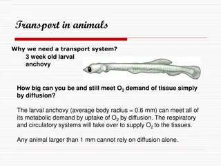
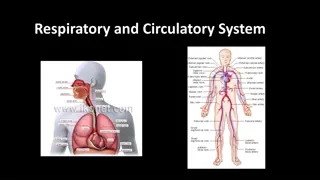
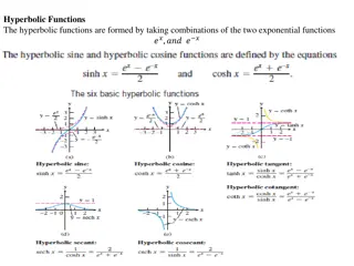
Understanding the Circulatory System: Functions and Components

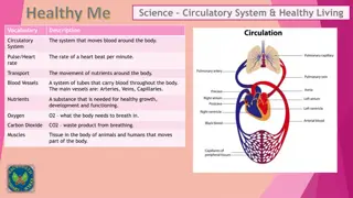
The circulatory system, comprised of pulmonary and systemic circulation, plays a crucial role in transporting oxygen, nutrients, and waste products throughout the body. It consists of the heart, arteries, veins, and capillaries which work together to ensure proper circulation. Blood, the carrier of essential substances, comprises plasma, red blood cells, white blood cells, and platelets. The heart, a central organ, pumps blood to all body cells, supporting various vital functions such as oxygen supply, waste removal, and hormone distribution.

Download Presentation

Please find below an Image/Link to download the presentation.
The content on the website is provided AS IS for your information and personal use only. It may not be sold, licensed, or shared on other websites without obtaining consent from the author. Download presentation by click this link. If you encounter any issues during the download, it is possible that the publisher has removed the file from their server.
E N D
Presentation Transcript
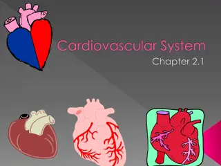
Circulatory System Ass.Prof.Dr. Suhad Faisal Hatem
The circulatory system actually consists of two separate systems: pulmonary circulation and systemic circulation The circulatory system is a transport system. important functions are: 1. Supplying oxygen from the lungs to the tissues 2.Supplying substances absorbed from the digestive system to the tissues 3.Removing carbon dioxide from the tissues to the lungs. 4.Removing waste products from the tissues to the kidneys. 5.Regulating body temperature increasing the diameter of blood vessels. increases heat loss, whereas reducing the diameter of the blood vessels prevents heat loss. 7.Distributing hormones and other chemicals to different parts of the body
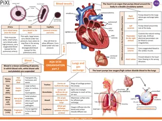
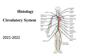
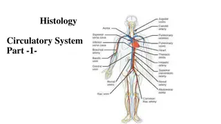
The system is made up of the heart , which functions as a pump and the vessels which act as channels through which blood is pumped. Arteries are blood vessels which carry oxygen-rich blood away from the heart to the various tissues in the body. The arteries of the body split up into successively narrow branches, until they form the very fine (microscopic) capillaries . Capillaries have very thin walls and allow the exchange of substances between body cells and the blood. Several capillaries join to form successively wider branches forming a vein . Veins carry oxygen-depleted blood back to the heart Blood
Blood is the carrier of oxygen and other substances. The normal total circulating volume is about 8% of the body weight (5.6 liters in a 70 kg man) Blood is a fluid connective tissue. It circulates throughout the body through blood vessels by the pumping action of the heart. Blood in arteries carries oxygen and nutrients to all the body s cells. Blood in veins carries carbon dioxide and other wastes away from the cells Blood also defends the body against infection, repairs body tissues, transports hormones, and controls the body s pH. Composition of Blood 1-Blood contains many dissolved substances and blood cells. Types of blood cells include red blood cells, white blood cells, and platelets. 2-Plasma has 90% water, 10% dissolved gases, salts, nutrients, enzymes, hormones, wastes, and proteins
Blood is made up of four major components: Blood Content 50% Water Plasma: the liquid portion. Contains dissolved nutrients, hormones, gases. 45% Red Blood cells Red blood cells: transport oxygen 4% Plasma with Substances White cells: defenses against invaders 1% Leukocytes (WBC) , Platelets Platelets: help form blood clots
HEART The heart is a muscular pear shaped organ It is protected by the bony rib cage, and its rarely damaged by a blow. It is located in the chest a little to the left of the center line. heart s function, like its position, is central to life. Without a continuous supply of blood (oxygen, sugar) the body cannot function. To maintain this supply, the heart has to work non-stop. The heart receives deoxygenated blood on the right side and pumps it to the lungs to be oxygenated. Oxygenated blood returns from the lungs to the left side of the heart, which pumps it to the rest of the body Deoxygenated Blood flows into the right upper chamber ( right atrium ) which is separated from the lower chamber ( ventricle ) by a one way valve. The deoxygenated blood is transported by two veins the superior and the inferior vena cava, bringing blood from the upper and lower halves of the body, respectively. When pressure in the atrium is greater than that in the ventricle, the valve opens, and blood flows from the atrium into the ventricle. The ventricle is filled with blood.
The valve closes once more and this marks the beginning of the contraction of the ventricle. The ventricle has very thick, powerful muscular walls. The right ventricle contracts, forcing blood out through the pulmonary artery , which also has a valve at its opening. The pulmonary artery takes the deoxygenated blood to the lungs, where it gets oxygenated once more. As most of the blood in the ventricle is pumped out the pressure in the ventricle drops, and the valve at the opening of the pulmonary artery closes. The ventricle relaxes.The cycle goes on as once more blood from the atrium enters the ventricle. aorta the main artery arising from the heart to be circulated throughout the body. The blood ultimately returns the right side of the heart in the deoxygenated condition.
Arteries Blood is ejected from the left ventricle through the aorta. This large artery (2.5 cm in diameter in the adult), has numerous branches, so that the oxygenated blood reaches the different parts of the body. Whenever blood flows through an artery, its flexible wall expands at each heartbeat and this can be felt as the pulse. The blood also exerts pressure on the vessel walls as it is pumped through. Pressure is necessary for circulation to take place. . The walls of arteries are lined with smooth cells (endothelium), around this is elastic tissue and muscle. The elastic and muscle layers make the arteries efficient in pumping blood through the body. One of the important factors which changes vessel diameters is the control by the nerves . Sympathetic nerves for example, simultaneously cause narrowing of blood vessels in the skin, while widening those in the muscles
Capillaries :- Most arteries divide as successively smaller branches, until they form very fine capillaries in all the body tissues. These are minute vessels with very thin walls. The thin walls allow for the movement of oxygen and other essential substances from the blood of the body cells. Waste material from the cell such as carbon dioxide diffuses from the cell to the capillary. Several capillaries join together to form a venule (a small vein), ultimately leading to a vein. If capillaries are cut blood oozes out, slowly.
Vein Once blood has passed through the capillary systems it must be returned to the heart. Done by veins Walls contains connective tissue and smooth muscle. Largest veins contain one way valves that keep blood flowing toward heart. Many found near skeletal muscles. When muscles contract, blood is forced through veins
BLOOD PRESSURE circulating blood presses against the walls of the blood vessels with a force/ pressure. The pressure is greatest during systole , when the heart contracts and forces blood into the arteries of the body. This maximum pressure is known as the systolic pressure . During the diastole , when the heart muscle relaxes, the pressure drops to a minimum. This is known as the diastolic pressure . It is influenced by the general health, heredity, age, activity, position of the body and emotional state. The blood pressure is determined by the cardiac output and the peripheral vascular resistance (i.e. the diameter of the blood vessels). Hence, if the heart pumps with more force and also at a greater rate, the cardiac output increases, increasing the blood pressure. if the arteries are more narrow, the pressure required to pump blood through them has to be more. During the stress response sympathetic nerves increase the rate and force of the heart beat and increase in blood pressure.
RECORDING THE BP Is done with a sphygmomanometer . It is recorded as the systolic pressure over the diastolic. A normal young adult may have a BP of 120/80 mmHg, this means that the systolic pressure is 120, while the diastolic is 80. Considerable variation within the normal range is possible. A systolic pressure above 140 mm, and a diastolic pressure above 90 mm, should be further investigated
PORTAL CIRCULATION Absorption of digested food materials takes place through the villi of the small intestine, into the capillaries. These capillaries join to form a portal vein, which transports the blood rich in absorbed substances, to the liver. Substances are stored in the liver. When required they are broken down, and enter the circulation via the hepatic vein. It joins the inferior vena cava, and hence transports blood to the right atrium
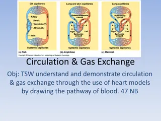
Summery Pulmonary circulation is the part of the circulatory system that carries blood between the heart and lungs Deoxygenated blood leaves the right ventricle through pulmonary arteries, which transport it to the lungs. In the lungs, the blood gives up carbon dioxide and picks up oxygen. The oxygenated blood then returns to the left atrium of the heart through pulmonary veins.
Systemic Circulation Systemic circulation is the part of the circulatory system that carries blood between the heart and body. Oxygenated blood leaves the left ventricle through the aorta. The aorta and other arteries transport the blood throughout the body, where it gives up oxygen and picks up carbon dioxide. The deoxygenated blood then returns to the right atrium through veins
Circulatory System Diseases Diseases of the heart and blood vessels called cardiovascular diseases (CVD), are very common. The leading cause of CVD is atherosclerosis. Atherosclerosis is the buildup of plaque inside arteries. it narrows the arteries and reduces blood flow and prevents blood circulation in the heart Plaque consists of cell debris, cholesterol, and other substances. . Stroke Atherosclerosis in the arteries of the brain can also lead to a stroke. A stroke is a loss of brain function due to a blockage of the blood supply to the brain. Risk factors for stroke include old age, high blood pressure, having a previous stroke, diabetes, high cholesterol, and smoking.
Coronary Heart Disease Atherosclerosis of arteries that supply the heart muscle is called coronary heart disease. blocked coronary artery can cause a heart attack. The loss of oxygen to the heart muscle cause that part of the tissue to die. A heart attack occurs when the blood supply to part of the heart muscle is blocked and cardiac muscle fibers die.