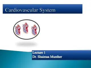
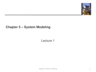
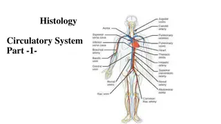
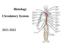
Understanding the Cardiovascular System: A Detailed Overview

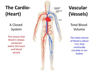
The cardiovascular system, comprising the heart, blood vessels, and blood, plays a crucial role in supplying fuel and oxygen to the body's cells while removing waste products. This system functions as a double pump, circulating blood through the pulmonary and systemic circulatory systems. Proper valve function is essential for efficient blood flow. Calculations of blood mass distribution provide insights into the body's overall circulation. Explore the intricacies of this vital system to understand its significance in maintaining overall health.

Download Presentation

Please find below an Image/Link to download the presentation.
The content on the website is provided AS IS for your information and personal use only. It may not be sold, licensed, or shared on other websites without obtaining consent from the author. Download presentation by click this link. If you encounter any issues during the download, it is possible that the publisher has removed the file from their server.
E N D
Presentation Transcript
Physics of the Physics of the Cardiovascular System Cardiovascular System
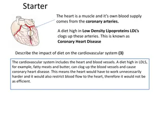
The cells of the body act like individual engines. In order for them to function they must have: - 1. Fuel from our food to supply energy. 2. O2 from the air we breathe to combine with the food to release energy. 3. A way to dispose of the by-products of the combustion (mostly CO2, H2O, and heat). Since the body has many billions of cells an elaborate transportation system is needed to deliver the fuel and O2 to the cells and remove the by-products. The blood performs this important body function. Blood represents about 7% of the body mass or about 4.5kg (~4.4 liters) in a 64kg person. The blood, blood vessels, and heart make up the cardiovascular system (CVS).
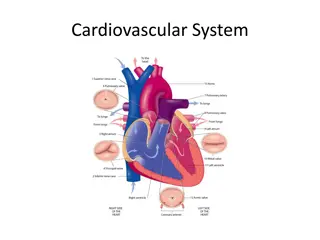
Major components of the cardiovascular system The heart is basically a double pump; it provides the force needed to circulate the blood through the two major circulatory systems: - The pulmonary circulation in the lungs. The systemic circulation in the rest of the body. The blood in a normal individual circulates through one system before being pumped by the other section of the heart to the second system.
. The heart has a system of valves that, if functioning properly, permit the blood to flow only in the correct direction. If these valves become diseased and do not open or close properly the pumping of the blood becomes inefficient The blood volume is not uniformly divided between the pulmonary and systemic circulation, but it is: Blood Circulation 100/100 Systemic Circulation 80/100 Arteries 15/100 Pulmonary Circulation 20/100 Arteries 46.5/100 Capillaries 10/100 Veins 75/100 Capillaries 7/100 Veins 46.5/100
Example: calculate the mass of the blood in all circulation of a person his body mass is 80Kg? Mass of Blood 80x7/100 = 5.6kg Mass of Blood in Systemic Circulation 5.6x80/100 = 4.48kg Arteries 4.48x15/100 = 0.672kg = 0.448kg Mass of Blood in Pulmonary Circulation 5.6x20/100 = 1.12kg Arteries 1.12x46.5/100 = 0.521kg = 0.078kg Capillaries 4.48x10/100 Veins Capillaries 1.12x7/100 Veins 4.48x75/100 = 3.360kg 1.12x46.5/100 = 0.521kg
Example: The mass of the pulmonary blood of a person is 1.5kg, find: - The mass of this person? The mass of his systemic blood? Pulmonary Mass = Blood Mass x 20/100 1.5 = Blood Mass x 20/100 Blood Mass = 7.5kg 1) The mass of this person: - Blood Mass = Body Mass x 7/100 7.5 = Body Mass x 7/100 Body Mass = 107kg 2) The mass of his systemic blood: - Systemic Mass = Blood Mass x 80/100 Systemic Mass = 7.5 x 80/100 Systemic Mass = 6kg
To the eye blood appears to be a red liquid slightly thicker than water. When examined by various physical techniques it is found to consist of several different components. The red color is caused by the red blood cells (erythrocytes). A nearly clear fluid called blood plasma accounts for the other 55%. The combination of red blood cells and plasma causes blood to have flow properties different from those of a fluid like water. Beside red blood cells and plasma, there are some important blood components, such as the white blood cells (leukocytes), present in small amounts. The blood also contains platelets. Platelets are involved in the clotting function of blood.
O2and CO2exchange in the capillary system Oxygen and carbon dioxide also diffuse through tissue. The most probable distance D that a molecule will travel after N collisions with other molecules with an average distance between collisions is: - D = N In tissue the density of the molecules is about 1000 times greater than in air; therefore, is much longer in air than in tissue. A typical value for in water, which can serve as a model for tissue, is about 10-11m, and a molecule makes about 1012collisions/sec. Thus after 1sec in water the most probable diffusion distance is about 10-5m or about a factor of 103less than in air. N D = 11 11 = = = 12 6 5 10 10 10 10 10 x m
This very short diffusion distance is the primary reason that the capillaries in tissue must be very close together. Not all capillaries are carrying blood at any one time. In resting muscle only 2 to 5% of the capillaries are functional Starling's law of capillary describes the flow of fluids into and out of the capillaries. Fluid movement through the capillary wall is the result of two pressures: - 1- The hydrostatic pressure across the capillary wall forcing fluids out of the capillary. 2- The osmotic pressure bringing fluids in.
Work done by the heart In a typical adult each contraction of the heart muscles forces about 80ml (about one-third of a cup) of blood through the lungs from the right ventricle and a similar volume to the systemic circulation from the left ventricle. In the process the heart does work. The pressures in the two pumps of the heart are not the same. In the pulmonary system the pressure is quite low because of the low resistance of the blood vessels in the lungs.
The 25mmHg, is about one-fifth of that in the systemic circulation. In order to circulate the blood through the much larger systemic network the left side of the heart must produce pressures that are typically about 120 mmHg at the peak (systole) of each cardiac cycle. During the resting phase (diastole) of the cardiac cycle the pressure is typically about 80mmHg. The muscle driving the left ventricle is about three times thicker than that of the right ventricle. In addition, the circular shape of the left ventricle is more efficient for producing high pressure than the elliptical shape of the right ventricle. maximum pressure (systole), typically about
The work done W by a pump working at a constant pressure P is equal to the product of the pressure and the volume pumped V, or W=P V. We can estimate the physical work done by the heart by multiplying its average pressure by the volume of blood that is pumped. Actually the pumping action takes place in less than one-third of the cardiac cycle and the heart muscle rests for over two-thirds of the cycle. * The heart rate of a person is 120 pulse/min; calculate the action time and the resting time of heart muscle. 120pulse/1min = 120pulse/60sec 2pulse/1sec = 1pulse/0.5sec 1pulse = 1/3contraction + 2/3 relaxation 0.5x1/3 = 0.17sec (the time of contraction) 0.5x2/3 = 0.33sec (the time of relaxation)
Pressure across the blood vessel wall (Transmural Pressure) The greatest pressure drop in the cardiovascular system occurs in the region of the arterioles and capillaries In order to understand why they do not burst we must discuss the law of Laplace, which tells us how the tension in the wall of a tube is related to the radius of the tube and the pressure inside the tube. Consider a long tube of radius R carrying blood at pressure P. We can calculate the tension T in the wall (T=RP).
Bernoulli's principle applied to the cardiovascular system Whenever there is a rapid flow of a fluid such as water, the pressure is reduced at the edge of the rapidly moving fluid. Bernoulli's principle is based on the law of conservation of energy. Pressure in a fluid is a form of potential energy PE since it has the ability to perform useful work. In a moving fluid there is kinetic energy KE due to the motion. This kinetic energy can be expressed as energy per unit volume such as ergs per cubic centimeter. If fluid is flowing through the frictionless tube, the velocity increases in the narrow section and the increased kinetic energy KE of the fluid is obtained by a reduction of the potential energy PE of the pressure in the tube.
As the velocity reduces again on the far side of the restriction the kinetic energy is converted back into potential energy and the pressure increases again as indicated on the manometers. How fast does your blood flow? As the blood goes from the aorta into the smaller arteries and arterioles with greater total cross-sectional areas the velocity of the blood decreases much as the velocity of a river decreases at a wide portion.
Notice that the blood velocity is related in an inverse way to the total cross-sectional area of the vessels carrying the blood. You are undoubtedly aware of the characteristic of a liquid called viscosity ( ). The viscosity of water is about 10-3Pas at 20oC [where Pas = 1 N s/m2= 1 kg/(m s) In the SI system ]. The viscosity of blood is typically 3x10⁻³to 4x10⁻³Pas but depends on the percentage of red blood cells in the blood. Poiseuille's law states that the flow through a given tube depends on the pressure difference from one end to the other (PA-PB), the length L of the tube, the radius R of the tube, and the viscosity of the fluid. When all of these variables are put together with a constant to keep the units working correctly we get Poiseuille's equation: - 1 In SI units the flow rate will be in cubic meters per second if PA-PBis in newtons per square meter, is in Pas, and R and L are in meters.
Blood flow-laminar and turbulent You have probably seen both a slow, smooth, quietly flowing river and a rapid, turbulent, noisy river. . The first type of river is analogous to the laminar or streamline flow that is present in most blood vessels. . The second is similar to the turbulent flow found at a few places in the circulatory system, for example, where the blood is flowing rapidly pass the heart valves. An important characteristic of laminar flow is that it is silent. If all blood flow were laminar, information could not be obtained from the heart with a stethoscope. The heart sounds heard with a stethoscope are caused by turbulent flow. During a blood pressure measurement, the constriction produced by the pressure cuff on the arm produces turbulent flow and the resulting vibrations can be detected with a stethoscope on the brachial artery.
If you gradually increase the velocity of a fluid flowing in a tube by reducing the radius of the tube, it will reach a critical velocity Vc when laminar flow changes into turbulent flow. The critical velocity will be lower if there are restrictions or obstructions in the tube. Osborne Reynolds determined that the critical velocity is proportional to the viscosity of the fluid and is inversely proportional to the density of the fluid and the radius R of the tube, Vc=K / R. The constant of proportionality K is called the Reynold's number, and it is approximately equal to 1000 for many fluids, including blood, flowing in long straight tubes of constant diameter. If there are bends or obstructions the Reynold's number becomes much smaller.
Find the kinetic energy (KE) of 2gm of blood leaving aorta of radius 1.2cm.
Heart sounds The heart sounds heard with a stethoscope are caused by vibrations originating in the heart and the major vessels. The opening and closing of the heart valves contribute greatly to the heart sounds; turbulent flow occurs at these times and the vibrations produced are often in the audible range. The amount and quality of the sound heard depend on the design of the stethoscope as well as on its pressure on the chest, its location, the orientation of the body, and the phase of the breathing cycle. The physics of some cardiovascular diseases Because of the many physical aspects of the cardiovascular system, heart diseases often have a physical component. Many of these diseases, for example, increase the work load of the heart or reduce its ability to work at a normal rate.
The work done by the heart is roughly the tension of the heart muscle and times how long it acts. Anything that increases the muscle tension or how long it acts will increase the work load of the heart. For example, high blood pressure (hypertension) causes the muscle tension to increase in proportion to the pressure. A fast heart rate (tachycardia) increases the work load since the amount of time the heart muscles spend contracting increases. To reduce the work load of the heart, bed rest and O2therapy are prescribed. Giving O2increases the O2in the blood so that less blood must be pumped to the tissues.
Other heart diseases such Heart valve defects, types of the Heart valve defects: - Stenosis: - the valve does not open wide enough. In stenosis the work of the heart is increased because a large amount of work is done against the obstruction of the narrow opening, and the blood supply to the general circulation is reduced. Insufficiency: - the valve does not close well enough. In insufficiency some of the pumped blood flows back into the heart so that the volume of the circulated blood is reduced. Man-made device that has helped heart patients is the artificial heart valve.
The functions of the blood 1. Red blood cells carry oxygen from the lungs to the tissues and carbon dioxide from the tissues to the lungs. Blood plasma transports antibodies, which protect the body from infection, carries food substances absorbed from the intestines and takes waste products to the kidneys for excretion. It also transports hormones, secreted by endocrine glands, to their sites of action. Clotting factors circulating in the plasma prevent blood loss whenever blood vessels are damaged or broken. Blood also regulates the water content of tissue cells. Blood buffers, chemicals within the blood, maintain the correct acidity of body fluids. Enzymes may be transported by blood plasma, which also distributes the heat produced by metabolism. 2. 3. 4. 5. 6.


")


 of 2gm of blood")