Understanding Blood Cells and Their Functions in the Circulatory System

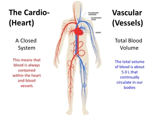
Blood is comprised of cells and plasma that circulate within the closed circulatory system. The formed elements include erythrocytes, platelets, and leukocytes, each playing vital roles in oxygen transport, defense against infection, and nutrient distribution throughout the body. The hematocrit estimates packed red blood cell volume, while plasma transports nutrients, metabolic residues, and hormones. Blood also regulates body temperature and acid-base balance. Explore the intricate components and functions of blood in this informative guide.
Download Presentation
Please find below an Image/Link to download the presentation.
The content on the website is provided AS IS for your information and personal use only. It may not be sold, licensed, or shared on other websites without obtaining consent from the author. Download presentation by click this link. If you encounter any issues during the download, it is possible that the publisher has removed the file from their server.

Presentation Transcript
Blood Cells: Introduction Blood consists of the cells and fluid that flow in a regular unidirectional movement within the closed circulatory system. made up of two parts: formed elements (blood cells), and plasma, the liquid in which the formed elements are suspended. The formed elements are erythrocytes (red blood cells), platelets, and leukocytes (white blood cells).
If blood is removed from the circulatory system, it will clot. This clot contains formed elements and a clear yellow liquid called serum, which separates from the coagulum. Blood that is collected and kept from coagulating by the addition of anticoagulants (e.g., heparin) separates, when centrifuged, into layers that reflect its heterogeneity. The hematocrit is an estimate of the volume of packed erythrocytes per unit volume of blood.
The translucent, yellowish, somewhat viscous supernatant obtained when whole blood is centrifuged is the plasma. The formed elements of the blood separate into two easily distinguishable layers. The lower layer represents 42 47% of the entire volume of blood in the hematocrit tube. It is red and is made up of erythrocytes. The layer immediately above (1% of the blood volume), which is white or grayish in color, is called the buffy coat and consists of leukocytes. These elements separate because the leukocytes are less dense than the erythrocytes. Covering the leukocytes is a fine layer of platelets not distinguishable by the naked eye.
functions Leukocytes, which have diversified functions are one of the body's chief defenses against infection. They circulate throughout the body via the blood vascular system, but while suspended in the blood they are round and inactive. Crossing the wall of venules and capillaries, these cells penetrate the tissues, where they display their defensive capabilities. The blood is a distributing vehicle, transporting oxygen, carbon dioxide (CO2), metabolites, and hormones, among other substances. O2is bound mainly to the hemoglobin of the erythrocytes, whereas CO2, in addition to being bound to the proteins of the erythrocytes (mainly hemoglobin), is carried in solution in the plasma as CO2or HCO3 .
The plasma transports nutrients from their site of absorption or synthesis, distributing them to various areas of the organism. It also transports metabolic residues, which are removed from the blood by the excretory organs. Blood, as the distributing vehicle for the hormones, permits the exchange of chemical messages between distant organs for normal cellular function. It further participates in the regulation of body temperature and in acid base and osmotic balance.
Cell Type Main Products Main Functions Erythrocyte Hemoglobin CO2and O2transport Leukocytes Specific granules and modified lysosomes (azurophilic granules) Phagocytosis of bacteria Neutrophil (terminal cell) Eosinophil (terminal cell) Specific granules, pharmacologically active substances Defense against parasitic helminths; modulation of inflammatory processes Basophil (terminal cell) Specific granules containing histamine and heparin Release of histamine and other inflammation mediators Monocyte (not terminal cell) Granules with lysosomal enzymes Generation of mononuclear-phagocyte system cells in tissues; phagocytosis and digestion of protozoa and virus and senescent cells B lymphocyte Immunoglobulins Generation of antibody-producing terminal cells (plasma cells) T lymphocyte Substances that kill cells. Substances that control the activity of other leukocytes (interleukins) Killing of virus-infected cells Natural killer cell (lacks T and B cell markers) Attacks virus-infected cells and cancer cells without previous stimulation Killing of some tumor and virus-infected cells Platelet Blood-clotting factors Clotting of blood
Composition of Plasma Plasma is an aqueous solution containing substances of low or high molecular weight that make up 10% of its volume. The plasma proteins account for 7% of the volume and the inorganic salts for 0.9%; the remainder of the 10% consists of several organic compounds eg, amino acids, vitamins, hormones, lipoproteins of various origins. Through the capillary walls, the low-molecular-weight components of plasma are in equilibrium with the interstitial fluid of the tissues. The composition of plasma is usually an indicator of the mean composition of the extracellular fluids in general. The main plasma proteins are albumin;,-, and -globulins; lipoproteins, and proteins that participate in blood coagulation, such as prothrombin and fibrinogen. Albumin, the most abundant component, has a fundamental role in maintaining the osmotic pressure of the blood.
Staining of Blood Cells Blood cells are generally studied in smears or films prepared by spreading a drop of blood in a thin layer on a microscope slide. The blood should be evenly distributed over the slide and allowed to dry rapidly in air. In such films the cells are clearly visible and distinct from one another. Their cytoplasm is spread out, facilitating observation of their nuclei and cytoplasmic organization. Blood smears are routinely stained with special mixtures of red (acidic) and blue (basic) dyes. These mixtures also contain azures, dyes that are useful in staining some structures of blood cells known as azurophilics (azure + Gr. philein, to love). Some of these special mixtures (eg, Giemsa, Wright's, Leishman's) are named for the investigators who introduced their own modifications into the original mixture.
Erythrocytes (red blood cells), which are anucleate, are packed with the O2-carrying protein hemoglobin. Under normal conditions, these corpuscles never leave the circulatory system. Most mammalian erythrocytes are biconcave disks without. When suspended in an isotonic medium, human erythrocytes are 7.5 m in diameter, 2.6 m thick at the rim, and 0.8 m thick in the center. The biconcave shape provides erythrocytes with a large surface-to-volume ratio, thus facilitating gas exchange ERYTHROCYTES
Leukocytes Leukocytes (white blood cells) migrate to the tissues, where they perform multiple functions and most die by apoptosis. According to the type of granules in their cytoplasm and the shape of their nuclei, leukocytes are divided into two groups: granulocytes (polymorphonuclear leukocytes) and agranulocytes (mononuclear leukocytes). Both granulocytes and agranulocytes are spherical while suspended in blood plasma, but some become ameboid after leaving the blood vessels and invading the tissues. Their estimated sizes refer to blood smears, in which the cells are spread and appear larger than they actually are in the blood.
Granulocytes possess two types of granules: 1. the specific granules that bind neutral, basic, or acidic components of the dye mixture and have specific functions and 2. azurophilic granules which stain purple and are lysosomes. Specific and azurophilic granules contain enzymes Granulocytes have nuclei with two or more lobes and include the neutrophils, eosinophils, and basophils.
All granulocytes are nondividing terminal cells with a life span of a few days, dying by apoptosis (programmed cell death) in the connective tissue. It is estimated that billions of neutrophils die by apoptosis each day in the adult human. The resulting cellular debris is removed by macrophages and does not elicit an inflammatory response. Being nondividing terminal cells, granulocytes do not synthesize much protein. Their Golgi complex and rough endoplasmic reticulum are poorly developed. They have few mitochondria (low energy metabolism) and depend more on glycolysis; they contain glycogen and can function in regions scarce in oxygen, such as inflamed areas. Agranulocytes do not have specific granules, but they do contain azurophilic granules (lysosomes) that bind the azure dyes of the stain. The nucleus is round or indented. This group includes lymphocytes and monocytes
LEUKOCYTES GRANULOCYTES AGRANULOCYTES Neutrophils Lymphocytes Eosinophils Monocytes Basophils
Leukocytes are involved in the cellular and humoral defense of the organism against foreign material. In suspension in the circulating blood, they are spherical, nonmotile cells, but they are capable of becoming flattened and motile on encountering a solid substrate. Leukocytes leave the venules and capillaries by passing between endothelial cells and penetrating the connective tissue by diapedesis (Gr. dia, through, + pedesis, to leap), a process that accounts for the unidirectional flow of granulocytes and monocytes from the blood to the tissues. (Lymphocytes recirculate.)
Diapedesis is increased in individuals infected by microorganisms. Inflamed areas release chemicals originating mainly from cells and microorganisms, which increase diapedesis. The attraction of specific cells by chemical mediators is called chemotaxis, a significant event in inflammation through which leukocytes rapidly concentrate in places where their defensive properties are needed. The number of leukocytes in the blood varies according to age, sex, and physiological conditions.
NEUTROPHILS Neutrophils constitute 60 70% of circulating leukocytes. They are 12 15 m in diameter (in blood smears), with a nucleus consisting of two to five (usually three) lobes linked by fine threads of chromatin
Immature neutrophils that have recently entered the blood circulation have a nonsegmented nucleus in the shape of a horseshoe (band forms). An increased number of band neutrophils in the blood indicates a higher production of neutrophils, probably in response to a bacterial infection. Neutrophils with more than five lobes are called hypersegmented and are typically old cells. Although the maturation of the neutrophil parallels the increase in the number of nuclear lobes under normal conditions, in some pathological conditions, young cells appear with five or more lobes. In females, the inactive X chromosome appears as a drumsticklike appendage on one of the lobes of the nucleus (Figure 12 5). However, this characteristic is not obvious in all neutrophils in a blood smear. The cytoplasm of the neutrophil contains two main types of granules. The more abundant granules are the specific granules, which are small, near the limit of resolution of the light microscope. The second granule population in neutrophils consists of azurophilic granules, which are lysosomes 0.5 m in diameter. Neutrophils also contain glycogen in their cytoplasm. Glycogen is broken down into glucose to yield energy via the glycolytic pathway of glucose oxidation. The citric acid cycle is less important, as might be expected in view of the paucity of mitochondria in these cells. The ability of neutrophils to survive in an anaerobic environment is highly advantageous, since they can kill bacteria and help clean up debris in poorly oxygenated regions, eg, inflamed or necrotic tissue. Neutrophils are short-lived cells with a half-life of 6 7 h in blood and a life span of 1 4 days in connective tissues, where they die by apoptosis. They are active phagocytes of bacteria and other small particles. Neutrophils are inactive and spherical while circulating but show an active ameboid movement upon adhering to a solid substrate, such as collagen in the extracellular matrix.
Neutrophils look for bacteria to engulf by pseudopodia and internalize them in vacuoles called phagosomes, whose membrane is derived from the neutrophil plasmalemma. Immediately thereafter, specific granules fuse with and discharge their contents into the phagosomes. By means of proton pumps in the phagosome membrane, the pH of the vacuole is lowered to about 5.0, a favorable pH for maximal activity of lysosomal enzymes. Azurophilic granules then discharge their enzymes into the acid environment, killing and digesting the microorganisms. During phagocytosis, a burst of O2consumption leads to the formation of superoxide (O22) anions and hydrogen peroxide (H2O2). O22is a short-lived free radical formed by the gain of one electron by O2. It is a highly reactive radical that kills microorganisms ingested by neutrophils. Together with myeloperoxidase and halide ions, it forms a powerful killing system. Other strong oxidizing agents (eg, hypochlorite) can inactivate proteins. Lysozyme has the function of specifically cleaving a bond in the peptidoglycan that forms the cell wall of some gram-positive bacteria, thus causing their death. Lactoferrin avidly binds iron; because iron is a crucial element in bacterial nutrition, lack of its availability leads to bacterial death. The acid environment of phagocytic vacuoles can itself cause the death of certain microorganisms. A combination of these mechanisms will kill most microorganisms, which are then digested by lysosomal enzymes. Dead neutrophils, bacteria, semidigested material, and tissue fluid form a viscous, usually yellow collection of fluid called pus. Several neutrophil hereditary dysfunctions have been described. In one of them, actin does not polymerize normally, and the neutrophils are sluggish. In another, there is a failure to produce O22, H2O2, and hypochlorite, and microbial killing power is reduced. This dysfunction results from a deficiency of NADPH oxidase, leading to a deficient respiratory burst. Children with these dysfunctions are subject to persistent bacterial infections. More severe infections result when neutrophil dysfunction and macrophage dysfunction occur simultaneously.
EOSINOPHILS Eosinophils are far less numerous than neutrophils, constituting only 2 4% of leukocytes in normal blood. In blood smears, this cell is about the same size as a neutrophil and contains a characteristic bilobed nucleus The main identifying characteristic is the presence of many large and elongated refractile specific granules (about 200 per cell) that are stained by eosin. Eosinophilic specific granules have a crystalline core (internum) that lies parallel to the long axis of the granule . It contains a protein called the major basic protein with a large number of arginine residues. This protein constitutes 50% of the total granule protein and accounts for the eosinophilia of these granules. The major basic protein also seems to function in the killing of parasitic worms such as schistosomes. The less dense material surrounding the internum is known as the externum, or matrix
An increase in the number of eosinophils in blood (eosinophilia) is associated with allergic reactions and helminthic (parasitic) infections. In tissues, eosinophils are found in the connective tissues underlying epithelia of the bronchi, gastrointestinal tract, uterus, and vagina, and surrounding the parasitic worms. In addition, these cells produce substances that modulate inflammation by inactivating the leukotrienes and histamine produced by other cells. They also phagocytose antigen antibody complexes. Corticosteroids (hormones from the adrenal cortex) produce a rapid decrease in the number of blood eosinophils, probably by interfering with their release from the bone marrow into the bloodstream.
BASOPHILS Basophils make up less than 1% of blood leukocytes and are therefore difficult to find in smears of normal blood. They are about 12 15 m in diameter. The nucleus is divided into irregular lobes, but the overlying specific granules usually obscure the division. The specific granules (0.5 m in diameter) stain metachromatically (change the color of the stain used) with the basic dye of the usual blood stains . This metachromasia is due to the presence of heparin. Specific granules in basophils are fewer and more irregular in size and shape than the granules of the other granulocytes (Figure 12 13). Basophilic specific granules contain heparin and histamine. Basophils may supplement the functions of mast cells in immediate hypersensitivity reactions by migrating (under special circumstances) into connective tissues There is some similarity between granules of basophils and those of mast cells. Both are metachromatic and contain heparin and histamine. Metachromasia is a property that enables certain substances to change the color of some basic dyes (eg, toluidine blue), being stained by the changed color (purple, in this example). Basophils can liberate their granule content in response to certain antigens, as can mast cells (see Chapter 5: Connective Tissue and Chapter 14: Lymphoid Organs). Despite the similarities they present, mast cells and basophils are not the same, for even in the same species they have different structures, and they originate from different stem cells in the bone marrow
LYMPHOCYTES Lymphocytes constitute a family of spherical cells with similar morphological characteristics. They can be classified into several groups according to distinctive surface molecules (markers), which can be distinguished by immunocytochemical methods. They also have diverse functional roles, all related to immune reactions in defending against invading microorganisms, foreign macromolecules, and cancer cells. Lymphocytes with diameters of 6 8 m are known as small lymphocytes. A small number of medium-sized lymphocytes and large lymphocytes with diameters up to 18 m are present in the circulating blood. This difference has functional significance in that some larger lymphocytes are believed to be cells activated by specific antigens. The small lymphocyte, which is predominant in the blood, has a spherical nucleus, sometimes with an indentation. Its chromatin is condensed and appears as coarse clumps, so that the nucleus is intensely stained in the usual preparations, a characteristic that facilitates identification of the lymphocyte.
In blood smears, the nucleolus of the lymphocyte is not visible, but it can be demonstrated by special staining techniques and with the electron microscope. The cytoplasm of the small lymphocyte is scanty, and in blood smears it appears as a thin rim around the nucleus. It is slightly basophilic, assuming a light blue color in stained smears. It may contain a few azurophilic granules. The cytoplasm of the small lymphocyte has a few mitochondria and a small Golgi complex; it contains free polyribosomes Lymphocytes vary in life span; some live only a few days, and others survive in the circulating blood for many years. Lymphocytes are the only type of leukocytes that return from the tissues back to the blood, after diapedesis.
Monocytes Monocytes are bone marrow-derived agranulocytes with diameters varying from 12 to 20 m. The nucleus is oval, horseshoe, or kidney shaped and is generally eccentrically placed. The chromatin is less condensed than that in lymphocytes. Because of their delicate chromatin distribution, the nuclei of monocytes stain lighter than do those of large lymphocytes. The cytoplasm of the monocyte is basophilic and frequently contains very fine azurophilic granules (lysosomes), some of which are at the limit of the light microscope's resolution. These granules are distributed through the cytoplasm, giving it a bluish-gray color in stained smears. In the electron microscope, one or two nucleoli are seen in the nucleus, and a small quantity of rough endoplasmic reticulum, polyribosomes, and many small mitochondria is observed. A Golgi complex involved in the formation of the lysosomal granules is present in the cytoplasm. Many microvilli and pinocytotic vesicles are found at the cell surface . Blood monocytes are not terminal cells; rather, they are precursor cells of the mononuclear phagocyte system. After crossing venule or capillary walls and entering connective tissues, monocytes differentiate into macrophages
Platelets Blood platelets (thrombocytes) are nonnucleated, disklike cell fragments 2 4 m in diameter. Platelets originate from the fragmentation of giant polyploid megakaryocytes that reside in the bone marrow. Platelets promote blood clotting and help repair gaps in the walls of blood vessels, preventing loss of blood. Normal platelet counts range from 200,000 to 400,000 per microliter of blood. Platelets have a life span of about 10 days. In stained blood smears, platelets often appear in clumps. Each platelet has a peripheral light blue- stained transparent zone, the hyalomere, and a central zone containing purple granules, called the granulomere. Platelets contain a system of channels, the open canalicular system, that connects to invaginations of the platelet plasma membrane. This arrangement is probably of functional significance in facilitating the liberation of active molecules stored in platelets. Around the periphery of the platelet lies a marginal bundle of microtubules; this bundle helps to maintain the platelet's ovoid shape. In the hyalomere, there are also a number of electron-dense irregular tubes known as the dense tubular system.
Actin and myosin molecules in the hyalomere can assemble to form a contractile system that functions in platelet movement and aggregation. A cell coat rich in glycosaminoglycans and glycoproteins, 15 20 nm thick, lies outside the plasmalemma and is involved in platelet adhesion. The central granulomere possesses a variety of membrane-bound granules and a sparse population of mitochondria and glycogen particles. Dense bodies (- granules), 250 300 nm in diameter, contain calcium ions, pyrophosphate, adenosine diphosphate (ADP), and adenosine triphosphate (ATP). These granules also take up and store serotonin (5-hydroxytryptamine) from the plasma. -granules are a little larger (300 500 nm in diameter) and contain fibrinogen, platelet-derived growth factor, and several other platelet-specific proteins. Small vesicles, 175 250 nm in diameter, have been shown to contain only lysosomal enzymes and have been termed -granules. Most of the azurophilic granules seen with the light microscope in the granulomere of platelets are - granules
Platelet Functions The role of platelets in controlling hemorrhage can be summarized as follows. Primary aggregation Discontinuities in the endothelium, produced by injuries, are followed by platelet aggregation to the exposed collagen, via collagen-binding protein in platelet membrane. Thus, a platelet plug is formed as a first step to stop bleeding. Secondary aggregation Platelets in the plug release an adhesive glycoprotein and ADP. Both are potent inducers of platelet aggregation, increasing the size of the platelet plug. Blood coagulation During platelet aggregation, factors from the blood plasma, damaged blood vessels, and platelets promote the sequential interaction (cascade) of approximately 13 plasma proteins, giving rise to a polymer, fibrin, that forms a three-dimensional network of fibers trapping red cells, leukocytes, and platelets to form a blood clot, or thrombus. Clot retraction The clot that initially bulges into the blood vessel lumen contracts because of the interaction of platelet actin, myosin, and ATP. Clot removal Protected by the clot, the vessel wall is restored by new tissue formation. The clot is then removed, mainly by the proteolytic enzyme plasmin, formed, through the activation of the plasma proenzyme plasminogen, by endothelium- produced plasminogen activators. Enzymes released from platelet -granules also contribute to clot removal.
Hemophilia A and B are clinically identical, differing only in the deficient factor. Both are due to sex-linked recessive inherited disorders. Blood from hemophiliac patients does not coagulate normally: the blood clotting time is prolonged. Persons with this disease bleed severely even after mild injuries, such as a skin cut, and may bleed to death after more severe injuries. The blood plasma of patients with hemophilia A is deficient in clotting factor VIII or contains a defective factor VIII, one of the plasma proteins involved in fibrin generation; in hemophilia B, the defect is in factor IX. In severe cases the blood is incoagulable. There are spontaneous hemorrhages in body cavities, such as major joints and the urinary tract. Generally, only males are affected by hemophilia A, because the recessive gene to factor VIII is in the X chromosome. Females may have one defective X chromosome, but the other one is usually normal. Females develop hemophilia only when they have the abnormal gene in both X chromosomes, a rare event. However, women with a defective X chromosome may transmit the disease to their male children.


,")