
Non Coronary Vascular Stents
Safe scanning of patients with non-coronary vascular stents (NCVS) in MRI settings in NHS Scotland. It covers key questions regarding the safety of patients with NCVS, including risks associated with MRI at different strengths, timing of MRI post-stent implantation, and the safety of various stent m
1 views • 9 slides

Automated CT Perfusion Imaging in Acute Ischemic Stroke: Overview
This presentation delves into the significance of automated CT perfusion imaging in diagnosing and treating acute ischemic stroke. It covers essential information such as the burden of stroke in Ontario, the critical aspect of time in stroke treatment, hyperacute stroke treatment goals, and the impo
5 views • 41 slides
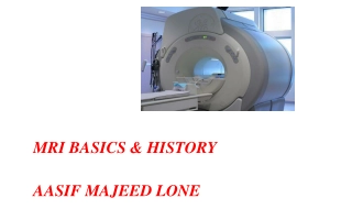
MRI BASICS & HISTORY AASIF MAJEED LONE
Magnetic Resonance Imaging (MRI) has revolutionized medical imaging, offering detailed views of the body without harmful radiation. This article delves into the historical development of MRI, highlighting key milestones such as the discoveries of the Rotating Magnetic Field by Nikola Tesla and the w
9 views • 49 slides

Premier Radiology: Quality Imaging Provider in Middle Tennessee
Premier Radiology, operating in Middle Tennessee under various names, offers a range of imaging services including X-Ray, CT, Mammography, and Ultrasound. The company does not plan to add MRI services to its Lebanon location but will acquire an existing unit. Detailed projections, utilization data,
0 views • 24 slides

Radiology Imaging Modalities for Renal System Evaluation
Various radiology modalities such as Ultrasound, X-Ray, MRI, CT, and Nuclear Scans are used to assess the renal system. Each modality has its advantages and disadvantages, with Ultrasound and MRI being radiation-free options, and Nuclear Scans providing functional assessment. Understanding the key f
6 views • 24 slides
Comprehensive Overview of Brain Imaging Techniques and Anatomy
Explore the world of brain imaging with functional MRI, MRI techniques, brain anatomy, neuronal activation, and brain vasculature explained in detail, shedding light on brain regions and their functions.
10 views • 53 slides

Basic Principles of MRI Imaging
MRI, or Magnetic Resonance Imaging, is a high-tech diagnostic imaging tool that uses magnetic fields, specific radio frequencies, and computer systems to produce cross-sectional images of the body. The components of an MRI system include the main magnet, gradient coils, radiofrequency coils, and the
4 views • 49 slides

Radiographic Imaging Methods of the Respiratory System
Radiographic imaging plays a crucial role in the evaluation and diagnosis of thoraco-mediastino-pleuro-pulmonary conditions. Techniques like radioscopy, digital radiography, computer tomography, magnetic resonance imaging, conventional pulmonary angiography, and hybrid imaging methods offer detailed
11 views • 21 slides

Radiological Investigation of Hepatobiliary System and Imaging Modalities Overview
Exploring the hepatobiliary system through radiology, this lecture covers the anatomy, modalities like X-ray, Ultrasound, CT scan, MRI, and nuclear scan used for imaging, their advantages, disadvantages, and indications. It delves into the significance of liver, gallbladder, and biliary duct imaging
2 views • 37 slides
Premier Radiology Mobile MRI Utilization Report
Premier Radiology, operating in Clarksville, ODC/MRICN2401-001, received 58 letters of support from various healthcare professionals, elected officials, and community members. They plan to open 18 diagnostic imaging centers, including a new facility in March 2024. The service area covers Montgomery
2 views • 20 slides

SALIVARY GLAND IMAGING
Salivary gland imaging plays a crucial role in diagnosing and monitoring diseases of the major salivary glands, including the parotid, submandibular, and sublingual glands. Different imaging techniques such as computed tomography, MRI, and ultrasound are used to visualize these glands and aid in dif
1 views • 12 slides

MRI Artifacts: Causes and Examples
MRI artifacts are signal misrepresentations that can be caused by various factors such as patient movement, magnetic field inhomogeneity, metallic objects, and under-sampling. Motion artifacts, magnetic susceptibility artifacts, and aliasing/wrap-around artifacts are common types of artifacts that c
2 views • 7 slides
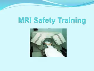
Importance of MRI Safety Precautions: Protecting Personnel and Participants
MRI safety is crucial to prevent severe injuries when working with the powerful magnetic fields generated by MRI scanners. This article covers safety training steps, badge access requirements, characteristics of magnetic fields, and the dangers associated with approaching an MRI scanner without prop
0 views • 31 slides
Advanced Imaging Technologies in Healthcare
Explore the world of digital imaging equipment including computed tomography (CT) scanners and magnetic resonance imaging (MRI) machines. Discover how these technologies work, their uses in medical diagnosis, and important facts to consider. Learn about the risks associated with CT scans and the ben
0 views • 59 slides
Diagnostic Reference Levels in Medical Imaging and Radiation Protection
The concept of Diagnostic Reference Levels (DRLs) is crucial in evaluating the amount of ionizing radiation used in medical imaging procedures. DRLs help determine if radiation levels are appropriate and need optimization. Authorized bodies establish numerical DRL values as advisory guidelines. Loca
1 views • 26 slides

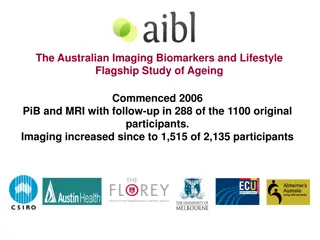
Australian Imaging Biomarkers and Lifestyle Study: Progress and Focus
The Australian Imaging Biomarkers and Lifestyle Study of Ageing (AIBL) commenced in late 2006, with a cohort size and follow-up progress detailed. The study includes assessments and biomarker imaging, such as MRI, amyloid PET, and tau PET scans, with ongoing review cycles and data additions. Current
0 views • 12 slides
Enhancing Stroke Treatment Response Through Advanced Imaging Management Center Strategy
This information details the strategy proposed by the Imaging Management Center to improve stroke treatment response. It emphasizes the importance of timely uploading of brain imaging data within specified timeframes using the AMBRA platform. The process involves modalities like NCCT, CTA, MRI, and
0 views • 6 slides

Comprehensive Overview of Jaw Imaging Modalities and Pathologies
Comprehensive overview detailing jaw imaging modalities such as X-Ray, CT, and MRI for evaluating jaw bone and tooth conditions. Covers anatomy, common mandibular lesions, odontogenic cysts, non-odontogenic cysts, and odontogenic benign tumors. Provides details on diagnostic protocols, imaging findi
0 views • 7 slides
Sparse Millimeter-Wave Imaging Using Compressed Sensing and Point Spread Function Calibration
A novel indoor millimeter-wave imaging system based on sparsity estimated compressed sensing and calibrated point spread function is introduced. The system utilizes a unique calibration procedure to process the point spread function acquired from measuring a suspended point scatterer. By estimating
2 views • 26 slides

Portal Vein Imaging Techniques and Anomalies Overview
Portal vein imaging is crucial for evaluating conditions affecting the abdominal part of the gastrointestinal tract. Techniques such as spleno-portography and CT triphasic contrast imaging are used to visualize the portal vein and diagnose anomalies like portal-systemic collaterals and porto-systemi
4 views • 7 slides
Insights into Structured Reporting Practices in Colorectal Cancer Imaging
A survey conducted by Dr. Eric Loveday at North Bristol NHS Trust revealed the current landscape of structured reporting in MRI and CT scans for rectal and colon cancer. Results indicate a positive outlook towards implementing national standards for structured radiology reporting, with an emphasis o
0 views • 7 slides

Introduction to Radiology: Imaging Modalities and Techniques
Radiology is a medical specialty that utilizes various imaging modalities such as X-Ray, MRI, CT, and Ultrasound to diagnose and treat patients. This field involves the supervision, performance, and interpretation of imaging studies, with findings reported to referring physicians. Radiology also inv
0 views • 10 slides
Peritoneal Pearls in Imaging: Case Study Analysis
A 45-year-old male presented with incidental lesions in the anterior peritoneal cavity during a CT scan for haematuria. These lesions, measuring between 9 mm and 22 mm, were non-enhancing and lacked consistent signal characteristics. Further imaging with MRI was recommended for better evaluation. By
0 views • 24 slides
Cutting-Edge iCMR Workshop Highlights and Participation Call
Dive into the latest innovations in interventional MRI (iCMR) with a series of hands-on workshops, informative sessions, and valuable opportunities to engage with experts in the field. Explore topics such as MRI catheterization, real-time MRI tutorials, and configuring iCMR suites. Learn about NHLBI
0 views • 7 slides
PowerGrid: Reconstructing High-Resolution Non-Cartesian MRI Data for Bioengineering Research
PowerGrid is a cutting-edge system developed by Brad Sutton, Assoc. Prof. at the University of Illinois, for harnessing high-performance computing to reconstruct large sets of high-resolution non-Cartesian MRI data. This technology addresses the need for improved image reconstruction packages in MRI
0 views • 10 slides
Australian Imaging Biomarkers and Lifestyle Study Overview
The Australian Imaging Biomarkers and Lifestyle Flagship Study of Ageing began in 2006 with PiB and MRI assessments. Over time, the study has expanded to include more participants and various imaging modalities. Current focuses include converting amyloid results to centiloid units, exploring blood b
1 views • 7 slides
Advanced Microscopy Techniques in EUV Lithography: SHARP Overview
SHARP utilizes Fresnel zone plate lenses to achieve diffraction-limited quality in EUV lithography, offering a range of NA values and image magnifications. The system allows emulation of mask-side imaging conditions with hundreds of lenses available. Coherence control and engineering are provided th
2 views • 18 slides

Methods of Imaging the Respiratory System
This lecture discusses various methods of imaging the respiratory system, including plain films, radionuclide imaging, CT scans, ultrasound for pleural disease, MRI, and bronchography. The normal anatomy relevant to respiratory system angiography is also highlighted through a series of images.
0 views • 27 slides
Insights into Multi-View Imaging System Optimization
Delve into the simulation and calibration of a multi-view imaging system using differentiable ray tracing and gradient-based optimization. Explore the challenges of ambiguity in results and the impact of angular offset on imaging accuracy. Discover how the system handles errors and maintains precise
0 views • 6 slides
Methods of Imaging the Brain: X-ray, CT Scan, and MRI
Different methods of imaging the brain, including X-ray, CT scan, and MRI, offer non-invasive ways to study brain structure and function. X-rays measure tissue density, CT scans provide detailed cross-sectional images, and MRI produces 3D anatomical images. MRI, in particular, has become a vital too
0 views • 37 slides
MRI Machine Safety and Considerations
MRI machines utilize powerful magnets and advanced technology for medical imaging. The magnets are superconducting, requiring ultra-cold temperatures for operation, and safety considerations include managing biological effects, area effects, and cryogenic gases. The magnetic fields used in MRI scans
0 views • 20 slides

Advanced MRI Protocols for Neuroimaging Studies
Cutting-edge MRI protocols, ADNI 3, aim to enhance brain imaging techniques for clinical trials, focusing on precise measurements, disease detection, and potential treatment evaluation by leveraging advanced imaging methods like ASL, dMRI, and TF-fMRI.
0 views • 20 slides
Magnetic Resonance Imaging (MRI)
Magnetic Resonance Imaging (MRI) is an imaging technique based on nuclear magnetic resonance principles. It was first developed in the 1970s by Paul Lauterbur and Peter Mansfield. MRI uses the interaction between protons in the body and magnetic fields to create detailed images. This technology has
0 views • 77 slides
The Menisci of the Knee Joint
Magnetic Resonance Imaging (MRI) is crucial in assessing knee joint internal derangements, including the menisci. These C-shaped structures play a vital role in providing stability, distributing weight-bearing strain, and facilitating joint movement. The medial and lateral menisci have distinct atta
0 views • 16 slides
Overview of DICOM WG21 Multi-Energy Imaging Supplement
The DICOM WG21 Multi-Energy Imaging Supplement aims to address the challenges and opportunities in multi-energy imaging technologies, providing a comprehensive overview of imaging techniques, use cases, objectives, and potential clinical applications. The supplement discusses the definition of multi
0 views • 33 slides
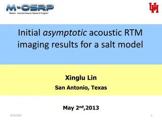
Initial Asymptotic Acoustic RTM Imaging Results in Salt Model
Acquire insights into the initial asymptotic acoustic RTM imaging results for a salt model in Xinglu Lin, San Antonio. This study delves into the concept of Reverse Time Migration (RTM), showcasing the methodology, workflow, and imaging conditions involved in this innovative seismic imaging techniqu
1 views • 22 slides
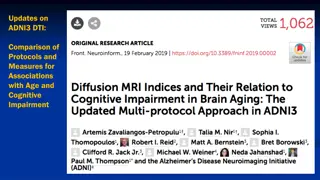
Insights into DTI and Advanced MRI Measures for Cognitive Impairment
Comparison of DTI protocols and measures in 317 participants revealed strong associations with age and cognitive impairment. MD and TDF-FA emerged as best measures, with robust connections to age. NODDI in advanced MRI allows for differentiation of A-beta positive and negative groups, offering great
1 views • 6 slides
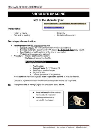
Comprehensive Guide to Shoulder MRI Imaging and Interpretation
Shoulder MRI imaging is valuable for assessing trauma, pain, swelling, deformity, and movement limitations. This detailed guide covers patient preparation, examination techniques, protocol, and interpretation of shoulder scans including assessment of tendons, ligaments, bursae, and bones.
0 views • 12 slides
Hip Imaging Basics: MRI Techniques and Sequences for Bilateral Examination
This content presents a quiz on hip imaging basics focusing on MRI techniques and sequences for bilateral examination. It covers topics such as appropriate positioning, the use of Propeller sequences, and the best choice of sequence for labral arthro-MR study. Images and questions related to hip ima
0 views • 15 slides
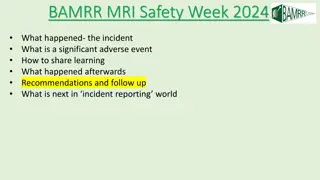
Insights from BAMRR MRI Safety Week 2024 Incident and Recommendations
The incident during BAMRR MRI Safety Week 2024 was more complex than initially thought, with numerous contributing factors identified. Immediate changes in practice were implemented, such as new procedures for general anesthesia, equipment checking, staff authorization, and physical safety measures.
0 views • 10 slides