Comprehensive Guide to Shoulder MRI Imaging and Interpretation
Shoulder MRI imaging is valuable for assessing trauma, pain, swelling, deformity, and movement limitations. This detailed guide covers patient preparation, examination techniques, protocol, and interpretation of shoulder scans including assessment of tendons, ligaments, bursae, and bones.
Download Presentation

Please find below an Image/Link to download the presentation.
The content on the website is provided AS IS for your information and personal use only. It may not be sold, licensed, or shared on other websites without obtaining consent from the author. Download presentation by click this link. If you encounter any issues during the download, it is possible that the publisher has removed the file from their server.
E N D
Presentation Transcript
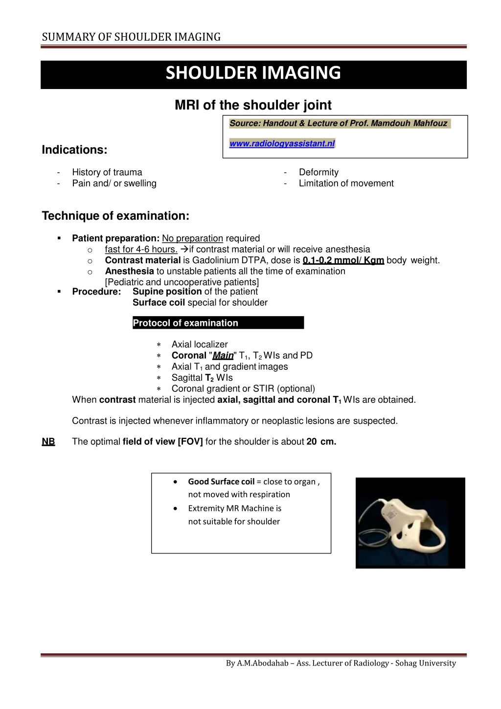
SUMMARY OF SHOULDERIMAGING SHOULDER IMAGING MRI of the shoulder joint Source: Handout & Lecture of Prof. Mamdouh Mahfouz www.radiologyassistant.nl Indications: - - History of trauma Pain and/ or swelling - - Deformity Limitation of movement Technique of examination: Patient preparation: No preparation required o fast for 4-6 hours. if contrast material or will receive anesthesia o Contrast material is Gadolinium DTPA, dose is 0.1-0.2 mmol/ Kgm body weight. o Anesthesia to unstable patients all the time of examination [Pediatric and uncooperative patients] Procedure: Supine position of the patient Surface coil special for shoulder Protocol of examination should include: Axial localizer Coronal "Main" T1, T2 WIs and PD Axial T1 and gradientimages Sagittal T2WIs Coronal gradient or STIR (optional) When contrast material is injected axial, sagittal and coronal T1 WIs are obtained. Contrast is injected whenever inflammatory or neoplastic lesions are suspected. The optimal field of view [FOV] for the shoulder is about 20 cm. NB Good Surface coil = close toorgan , not moved withrespiration Extremity MR Machine is not suitable for shoulder By A.M.Abodahab Ass. Lecturer of Radiology - Sohag University
SUMMARY OF SHOULDERIMAGING Landmark of Scan : o Axial Localizer o Identify "ICE CREAM CONE" = Main axis of scan Coronal Sagital Angle other than this = Scan Must be repeated Image interpretation [ Normal anatomy] Items to assessed: Supraspinatous, Infraspinatous, subscapularis, Biceps Anterior, Posterior, Superior, Inferior Superior, middle and inferior Glenohumeral ligaments Subacromial- subdeltoid, subcoracoid and subscapularis bursae Tendons Labrium Ligaments Bursae Acromion, acromio- clavicular joint, humeral head Bones Synovial effusion By A.M.Abodahab Ass. Lecturer of Radiology - Sohag University
SUMMARY OF SHOULDERIMAGING Tendons: All tendons appear normally black in all pulse sequences [Mature fibrous tissue contains non mobile H protons] Supraspinatous tendon is assessed in the coronal T1 and T2WIs First identify the acromio-clavicular joint then look underneath the joint you can see the black signal band inserting into the humeral greater tuberosity [In the same image you can see the subacromial bursa above the tendon however the bursa is not seen unless it is filled with fluid in cases of bursitis or rupture of the supraspinatous tendon] Intermediate signals may be seen within the tendon in T1 and PD images which are normal NB variants, these signal should not be normally seen in T2 WIs By A.M.Abodahab Ass. Lecturer of Radiology - Sohag University
SUMMARY OF SHOULDERIMAGING Infraspinatous tendon is also assessed in the coronal images underneath the spine of the scapula. The tendon also interests into the humeral greater tuberosity. Subscapularis tendon is best assessed in the axial image at the level of the bicipital groove The bicipital groove is a notch seen in the anterior aspect of the humeral head separating NB between the greater and lesser tuberosities. In Coronal Images it seen under coracoid. The subscapularis tendon appears as a thick low signal band stretched between the lesser tuberosity and the subscapularis muscle. In the same images if you look along the posterior aspect of the humeral head you will see the infraspinatous tendon [another site where you can evaluate the infraspinatous tendon] By A.M.Abodahab Ass. Lecturer of Radiology - Sohag University
SUMMARY OF SHOULDERIMAGING By A.M.Abodahab Ass. Lecturer of Radiology - Sohag University
SUMMARY OF SHOULDERIMAGING By A.M.Abodahab Ass. Lecturer of Radiology - Sohag University
SUMMARY OF SHOULDER IMAGING The biceps tendon appears as a small ball of low signal inside the bicipital groove in the axial images If you want to see the whole length of the biceps tendon go to the most anterior images of NB the coronal sections then you will seen the tendon in the bicipital groove crossing over the humeral head to insert into the superior labrium- glenoid complex. In the same image you can see the subscapularis tendon as well as the coracoid process. Underneath the coracoid process lies the subcoracoid bursa which is not usually identified unless filled with fluid. For better assessment of the biceps tendon, MR arthrography is usually indicated. Glenoid labrium: The labrium is triangular in shape, the base of the triangle is attached to the glenoid part of the scapula while the apex of the tringle points to the humeral head The labrium appears normally black in all pulse sequences [fibro-cartilagenous tissue contains non mobile H protons] The anterior and posterior labrium are assessed in the axial images [assess the anterior labrium in the same image where you assess the subscapularis tendon, not in any other plane] The superior and inferior labrium are assessed in the coronal plane Bursae: Normally the bursa is not seen unless filled with fluid [either due to synovial effusion, inflammation or after arthrography] Fluids appear of low signal in T1WIs and high signal in T2WIs [In cases of arthrography the injected contrast material will appear of high signal in T1 and T2 WIs] The subacromial bursa is seen in the coronal images above the supraspinatous tendon and below the acromio-clavicular joint "& extend below deltoid The subcorcoid bursa is seen in all images underneath the corcoid process [The subcorcoid bursa is usually continuous with the joint space, so you can see fluid in the bursa if there is synovial effusion in the joint, then do not diagnose bursitis in such case] The subscapularis bursa is located underneath the subscapularis tendon. This bursa is not frequently seen in the clinical practice. By A.M.Abodahab Ass. Lecturer of Radiology - Sohag University
SUMMARY OF SHOULDERIMAGING Bones [acromion, acromio-clavicular joint, humeral head] Acromion: The acromial shape is assessed only in the sagittal images. There are 4 types of acromion Type I with concave under surface. Type II with straight under surface. Type III with inferior hook. Type IV with convex under surface. Type I and II are normal shapes of the acromion while type III and IV are considered pathological. The acromial shape should be mentioned in the report whether normal or abnormal. NB Acromio- clavicular joint: best evaluated in the coronal and sagittal planes. The normal joint has a smooth upper and lower surfaces with no superior or inferior bulges. The acromion and clavicle should show normal marrow signal in all pulse sequences. Humeral head is assessed in all images specially the axial images. Normally it has a smooth cortical outline. The biceptal groove is seen in its anterior aspect. The humeral head usually show heterogenous marrow signal reflecting the presence of red and yellow marrow. The red marrow normally shows intermediate signal in all pulse sequences. NB By A.M.Abodahab Ass. Lecturer of Radiology - Sohag University
SUMMARY OF SHOULDERIMAGING A notch may be seen in the superior- lateral aspect of the humeral head near the insertion of NB the supraspinatous tendon in the coronal images [normal variant]. Synovial effusion: No fluids normally seen within the shoulder joint. When joint effusion is present, it is best assessed in the coronal T2 WIs filling the axillary pouch The axillary pouch is the space between the glenoid bone and humeral head in the inferior NB aspect of the shoulder joint. This pouch is formed by the inferior glenoid humeral ligament which is attached near the inferior labrium connecting the glenoid to the humeral head. The shoulder joint space is normally connected to NB The sheath of the biceps tendon. The subcoracoid bursa. So You should normally expect some fluid in these two anatomic sites when shoulder joint effusion is present. Glenohumeral ligaments [ superior, middle, inferior] All ligaments appear normally as low signal lines in all pulse sequences [mature fibrous tissue contains non mobile H protons] Inferior gleno- humeral ligament [IGHL] best evaluated in the coronal images forming the axillary pouch. The ligament is not usually discriminated from the inferior glenoid labrium (both appear low signal in all pulse sequences). Separation of the ligament from the labrium can be achived when joint effusion is present or after shoulder arthrography. Avulsion of the glenoid attachement of the IGHL= GAGL NB Avulsion of the humeral attachment of the IGHL = HAGL Middle gleno- humeral ligament [MGHL] best evaluated in the axial images. Normally it appears as a thin short line between the subscapularis tendon and the anterior glenoid labrium. NB By A.M.Abodahab Ass. Lecturer of Radiology - Sohag University
SUMMARY OF SHOULDER IMAGING This ligament may be normally absent in about 15% of cases Thickening of the ligament is considered normal variant and is known as "Buford complex" By A.M.Abodahab Ass. Lecturer of Radiology - Sohag University
SUMMARY OF SHOULDERIMAGING MR shoulder arthrography Is now considered the accurate method for evaluation of the shoulder anatomy and pathology. Many radiologists and clinicians believe that without arthrography, the conventional MR examination of the shoulder can miss a lot of abnormalities. Technique: 1 ml of Gd DTPA diluted in 20 ml normal saline injected into the shoulder joint space under screen control with the patient supine and his arm is slightly abducted and externally rotated [about 5- 10ml of the diluted contrast are injected, some iodinated contrast material like urographin is added to the solution so that you can see the fluid while injected under screen control] NB You should know that Gd- DTPA is not radioopaque?! Diagnosticvalue Normal anatomy: some of the anatomical structures are not easily evaluated without arthrography such as: All the gleno- humeral ligaments specially the superior one. The whole course of the biceps tendon The inferior labrium. Pathology: Many lesions require MR arthrography for adequate delineation and accurate assessment such as: All types of supraspinatous tendon tears specially the minor ones All types of anterior labral tears specially Persth's type, GLAD type and ALAPS type. All types of SLAP injuries except type I lesions All types of gleno- humeral ligament injuries including HAGL, GAGL, tears of the MGHL and SGHL By A.M.Abodahab Ass. Lecturer of Radiology - Sohag University
SUMMARY OF SHOULDERIMAGING By A.M.Abodahab Ass. Lecturer of Radiology - Sohag University
