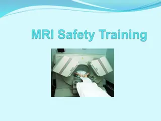
Importance of MRI Safety Precautions: Protecting Personnel and Participants
MRI safety is crucial to prevent severe injuries when working with the powerful magnetic fields generated by MRI scanners. This article covers safety training steps, badge access requirements, characteristics of magnetic fields, and the dangers associated with approaching an MRI scanner without proper precautions.
Download Presentation

Please find below an Image/Link to download the presentation.
The content on the website is provided AS IS for your information and personal use only. It may not be sold, licensed, or shared on other websites without obtaining consent from the author.If you encounter any issues during the download, it is possible that the publisher has removed the file from their server.
You are allowed to download the files provided on this website for personal or commercial use, subject to the condition that they are used lawfully. All files are the property of their respective owners.
The content on the website is provided AS IS for your information and personal use only. It may not be sold, licensed, or shared on other websites without obtaining consent from the author.
E N D
Presentation Transcript
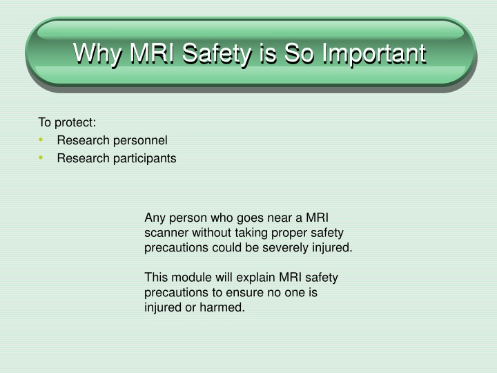
Why MRI Safety is So Important To protect: Research personnel Research participants Any person who goes near a MRI scanner without taking proper safety precautions could be severely injured. This module will explain MRI safety precautions to ensure no one is injured or harmed.
Safety Training MRI safety training consists of: Completing an MRI Screening Sheet Completion of MRI area walk-through with completion of checklist (if applicable) ID badge access-once all training is complete
Your MRI Badge Once you have completed all MRI Safety Training, you will be able to get badge access in order to work within the MRI scanner areas. After completion of all training you will need to email MRI Research Operations Manager, Jamie Weathersbee and include the following information: Full name and 9 digit # on the back of you badge Photo of the front and back of your badge (ID badge must be a valid UVA Health System Badge)
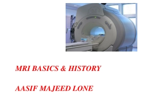
The MRI Scanner is Formidable The MRI Scanner is a very large and powerful magnet The MRI magnet is always ON at full power even when not actively scanning a patient Image Courtesy of Siemens Healthcare The force of the field is greatest at the periphery of the magnet. This force INCREASES as you move closer to the magnet
Characteristics of the Magnetic Field The force of the magnetic field is measured in tesla (T); a typical scanner is approximately 1.5- 3.0 tesla 3 tesla = 30,000 gauss For reference, the Earth s magnetic field is 0.5 gauss Five gauss and below are considered 'safe' levels of static magnetic field exposure for the general public Not all MRI magnets have the same field strength, thus their attractive forces may be different Fringe field The perimeter around a MR scanner within which the static magnetic fields are higher than five gauss
Fringe Field This line specifies the perimeter around a MR scanner within which the static magnetic fields are higher than five gauss As you approach the magnet, the fringe magnetic field gets STRONGER
Magnetic Field What objects can you take into a magnetic field? Work closely with the MRI Technologist who works in that type of environment each day. Anything that contains iron, cobalt or nickel must never be taken into the magnetic field.
MRI Sentinel Events The following may occur during the MRI scanning process when safety precautions are not taken: Missile effect or projectile injury from ferromagnetic objects Surgical implant injury from pacemakers, aneurysm clips, orbital metal Patient burns
Potential Projectiles Any ferromagnetic object may be attracted to the MRI scanner and become a projectile this is known as the missile effect The greater the amount of ferromagnetic material, the greater the force of attraction Remember, the magnetic field extends beyond the bore of the magnet in all directions (fringe field)
Potential Projectiles Small Objects Cell phone Keys Glasses Hair pins / barrettes Jewelry Safety pins Paper clips Coins Pens Pocket knife Nail clippers Steel-toed boots / shoes Tools Clipboards No loose metallic objects should be taken into the MRI Scan room!
Potential Projectiles Large Objects Due to the strength of the magnet, large objects such as chairs and IV poles can become projectiles and get stuck in the magnet! If a person is in the path of the object, serious injury could occur
Burns Caused by MRI It is possible for patients to get 1st, 2nd, or even 3rd degree burns in a MRI if items such as ECG cables are looped and are touching the patient s skin (even if these devices are MRI compatible) Any cable should not touch the patients skin directly, and should NOT be in a LOOPED configuration
Auditory Harm Activation of gradient magnetic fields produces significant vibrations in the gradient coils. MRI acoustical noise has been shown to produce reversible hearing impairment and could potentially produce permanent damage FDA Safety Guidelines for MR Devices Acoustic noise level International standard: 140 dB relative to 20 mPa Hearing protection is recommended for all patients undergoing a MRI procedure on a high-field MRI system (1.5T and 3.0T) Noise attenuating ear-plugs or head phones are routinely used in MRI
QUENCH Quench is done in an emergency to bring the magnetic field to ZERO in order to remove a patient from the scanner in extreme emergencies. MR scanners are super cooled with inert gases such as helium If these cryogens BOIL OFF either intentionally or unintentionally, a quench has occurred The issue with a quench is the potential for asphyxiation and frost-bite to the healthcare provider and patient If a quench occurs, remove all patients and staff from the room immediately The only time the magnet will be quenched on purpose is for a large fire, or is a staff member or research subject are pinned against the scanner.
Peripheral Nerve Stimulation (PNS) The rapid switching on and off of the magnetic field gradients is capable of causing nerve stimulation. Volunteers report a twitching sensation when exposed to rapidly switched fields, particularly in their extremities.
MRI Screening Sheet All research personnel are required to complete an MRI screening form to be reviewed by an MRI Technologist prior to working in the MRI area
Patient Screening and Contraindications NO ONE should enter the scan room without first being cleared by an MRI Technologist Some implants/devices are contraindicated for an MRI scan If a subject answers yes to any question on the MRI screening sheet, that issue must be addressed and resolved prior to entering the scan room NO defibrillators, aneurysm clips or electronic or magnetically activated devices Cardiac Pacemakers may be allowed after proper evaluation (patients only)
Participant Screening All research participants/subjects must complete a screening sheet prior to entering the MRI scanner room If someone has worked as a machinist, grinder, or welder and cannot absolutely confirm they always wore eye protection, they must first have orbital x-rays to confirm that there are no loose metallic bodies in the eye Any person who was injured by a metallic foreign body such as a bullet, BB, or shrapnel may not be able to proceed with an MRI scan unless there is proof that any remaining metal in the body is not in a location where it may move and cause injury/death
MRI Safety Zones There are 4 MRI Safety Zones: Zone 1 Zone 2 Zone 3 Zone 4
UVA MRI Safety Zones Zone 1 Zone 1: This is an uncontrolled, general access area It may be accessed by personnel without restrictions on materials or equipment in the area
UVA MRI Safety Zones Zone 2 Zone 2: This zone is the area between the general access zone, which is uncontrolled, and the strictly controlled zone III. Ferromagnetic items are safe in this zone but should not proceed beyond this zone unless needed for patient care.
UVA MRI Safety Zones Zone 3 Zone 3: This is a strictly controlled area. ID badge access is required to enter this zone. This zone is restricted to the general public or non-MRI Department trained personnel. Personnel in this zone must be screened and oriented to the MRI Department policy/procedures before entering.
UVA MRI Safety Zones Zone 4 Zone 4: This zone is located inside of Zone 3. This area is where the MRI Magnet resides (MRI Scanning Room) This area is strictly controlled and restricted to properly MRI safety training personnel Ferrous detectors are set up at entry into the scan room as well for enhanced safety NO ferromagnetic items are authorized in Zone 4
MRI Compatible Equipment All equipment must be tested for MR safe, unsafe, or conditional status. The internal components of electronic equipment can be damaged if exposed to a high magnetic field if not designed and built specifically to work in the MRI environment. ALL equipment including passive devices such as chairs and tables will be tested and marked with one of the following labels as approved by the American Society of Testing and Materials (ASTM). MRI Safe MRI Conditional Not Safe for MRI
MRI Compatible Equipment MRI SAFE: Objects/equipment with this label must be completely non-metallic, non-conductive, and non-ferromagnetic. These objects have no restrictions as to their use and location in the MRI environment. MRI CONDITIONAL: Objects with this label are known to cause no hazards in the MRI environment UNDER SPECIFIC CONDITIONS OF USE. These objects may remain in the presence of the MR scanner, but may have restrictions as to how close they may be, or operational settings at the time of image acquisition. The position and status of all MRI conditional devices must be confirmed by the MRI safety officer to ensure that the MRI conditions for each device are met. MRI UNSAFE: These items are considered unsafe in the MRI environment under any conditions. They must remain behind the 5 gauss line AT ALL TIMES.
Emergency Situations In the event of an emergency, first remove the patient from the MRI scan room Stand near the doors to the scan room to ensure no unauthorized emergency personnel can enter
Emergency Situations The MRI department has MRI conditional fire extinguishers accessible in zone II and zone III. It works the same way as other fire extinguishers: Pull Aim Squeeze Sweep
Other Information Radiology and Medical Imaging Core Website: https://med.Virginia.edu/radiology-research/ For scheduling related questions please contact our Administrative Coordinator, Jessica Lilly at: JLW8FK@hscmail.mcc.virginia.edu or 434-982-2585, or email RadiologyImagingCore@virginia.edu MRI Operations Manager: Jamie Weathersbee JG6W@hscmail.mcc.virginia.edu 434-243-1674
