Understanding the Meninges, Ventricles, and CSF in the Central Nervous System


Meninges, Ventricles, and CSF
Meninges, Ventricles, and CSF


Objectives
At the end of the lecture, the students should be able to:
Explain the
cerebral meninges
& compare between the main
dural folds
.
Identify the
spinal meninges
& locate the level of the
termination
of each of
them.
Describe the importance of the
subarachnoid space
.
Explain the
ventricular system
of the CNS and locate the site of each of
them.
Analyze the
formation
,
circulation
,
drainage
, and
functions
of the CSF.
Justify the
clinical point
related to the CSF.


M
e
n
i
n
g
e
s
1- (The dura surrounds
the brain and the spinal
cord and is responsible
for keeping in the CSF.)
2- (from which it is separated by
the subarachnoid space .The
delicate arachnoid layer is
attached to the inside of the dur
and surrounds the brain and
spinal cord.)
3- (Pia mater is a
thin fibrous tissue that
is impermeable to fluid.
This allows the pia
mater to enclose csf)

02:02
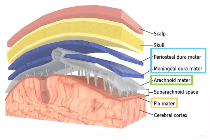
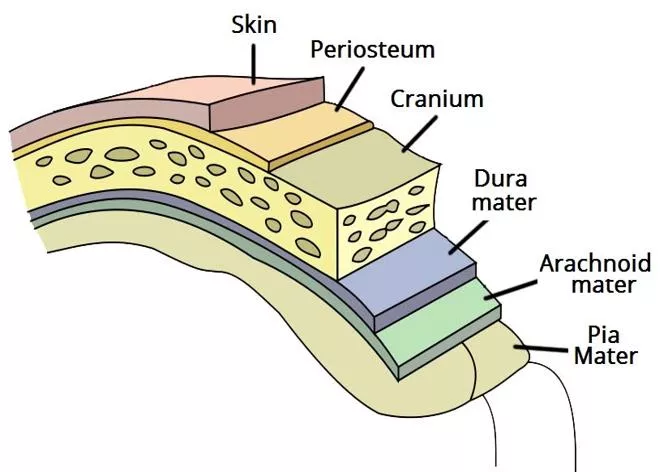
The brain and spinal cord
(CNS)
are invested by
three
concentric membranes/
layers
:
1-The outermost layer is the
dura
matter.
2-The middle layer is the
arachnoid
matter.
3-The innermost layer is the
pia
matter.
D
u
ra
O
u
tside
P
i
a
I
nside
o
The
cranial dura
is a two layered
tough
,
fibrous
,
thick membrane that surrounds the brain.
o
It is formed of two layers;
1)
periosteal
: attached to the skull.
2)
meningeal
:
folded forming the dural folds
*
falx
cerebri
, and
tentoriam cerebelli
.
o
Sensory innervation
of the dura is mostly from the
three meningeal branches of:
•
the
trigeminal
•
vagus
nerves
•
C1 to C3
(upper cervical nerves).
Meninges
1
-
D
u
r
a
M
a
t
t
e
r
*protect the brain
2. Tentorium cerebelli;
o
A
horizontal
shelf of dura, lies between the posterior part
of the
cerebral hemispheres
and the
cerebellum
.
o
It has a
free
border that encircles the midbrain.
o
Its superior surface in the middle line it is continuous
(attached)
with the falx cerebri, separated by the
straight sinus .
1. Falx cerebri;
o
It is a
vertical
sickle shaped sheet of dura, in the midline
o
Extends from the cranial roof into the
great longitudinal
fissure
between the two cerebral hemispheres.
o
It has an
attached
border adherent to the skull
o
And a
free
border lies above the
corpus callosum
Meninges
1
-
D
u
r
a
M
a
t
t
e
r
Two large reflection of dura extend into the cranial cavity;
Meninges
2
-
A
r
a
c
h
n
o
i
d
M
a
t
e
r
3
-
P
i
a
M
a
t
e
r
o
is a soft,
translucent
membrane
loosely
envelops the brain.
o
It is separated from the dura by a narrow
subdural
space.
o
is the innermost,
thin
, delicate &
highly
vascular
membrane that is closely
adherent
(attached)
to the gyri and fitted into the sulci.
o
Between the pia and arachnoid mater lies the
subarachnoid
space
which contains; fibrous
trabeculae, main blood vessels and CSF.
1- The
cisterna magna
, or cerebllomedullary cistern
o
lies between the inferior surface of the
cerebellum and the back of the medulla.
o
from this cistern CSF flows
out
of the fourth
ventricle.
2- Interpeduncular cistern
;
o
Is located at the
base
of the brain, where the
arachnoid spans the space between the two
cerebral peduncles of midbrain.
o
It contains the optic chiasma & circulus arteriosus
of Wills
and CSF
.
Meninges
S
u
b
a
r
a
c
h
n
o
i
d
S
p
a
c
e
The subarachnoid space is varied in depth forming;
subarachnoid cisterns.
Just like the brain
the spinal cord, is invested by
three
meningeal coverings:
the
pia
mater
, arachnoid
mater and
dura
mater.
Pia mater,
o
Innermost covering, a delicate membrane closely envelops the
cord and nerve roots.
o
It is attached through the arachnoid to the dura by the
denticulate
ligament
.
*
Dura mater;
o
the outer covering, is a single, tough fibrous membrane.
o
It envelopes the cord loosely .
o
It is separated from arachnoid matter by the
subdural space
, and
from the bony wall of the vertebral canal by the
epidural space
.
Arachnoid matter;
o
is a translucent membrane, lies between the pia and dura,
o
Between it and pia lies the subarachnoid space
contains CSF
.
S
p
i
n
a
l
M
e
n
i
n
g
e
s
The spinal meninges are very similar to the cranial meninges with
2 differences: 1) the epidural space and 2) denticulate ligament
*The denticulate ligament (pia) passes through the arachnoid and attaches to the dura
o
Spinal cord
terminates at level
L1-L2
.
o
Arachnoid
and
dural
and,
subarachnoid
space
, continue caudally to
S2
.
o
Pia
extends downwards forming the
filum
terminalis
which pierces the arachnoid and
dural sacs and passes through the
sacral hiatus
to be attached to the back of the
coccyx
.
S
p
i
n
a
l
M
e
n
i
n
g
e
s
Dr. Sanaa: Lumbar puncture is done between L3 and L4
Important!
V
e
n
t
r
i
c
u
l
a
r
S
y
s
t
e
m
o
Interconnecting channels within the CNS.
•
In the
spinal cord
; represented by the
central canal
.
•
Within the
brain
; a system of
4
ventricles is found.
o
The central canal of the spinal cord is
continuous upwards
to the
fourth ventricle
.
o
On each side of the fourth ventricle laterally, lateral recess extend to
open into
lateral aperture
opening
(foramen of
Luscka
), central defect in its roof (foramen of
Magendie
)*
o
The forth ventricle is
continuous up with
the
cerebral aqueduct
, that
opens in
the
third ventricle
.
o
The third ventricle is
continuous with
the
lateral ventricle
through the
interventricular foramen
(foramen of monro).
(brain and spinal cord)
*in the fourth ventricle there are 2 lateral recess which have an opening called foramen of luscka, there is another opening on
the wall called foramen of magendie. These openings allow CSF to pass from the ventricular system to the subarachnoid space
o
Present
in
the
ventricular system
, together with the
cranial
and
spinal subarachnoid spaces
.
o
It is
colourless fluid
containing little protein and few
cells.
o
It is about 150 ml.
(125 – 150)
o
It serves to
cushion
the brain from sudden
movements of the head
(protects brain and spinal cord)
o
It is
produced by the choroid plexus
(cluster of capillaries)
,
which is
located in the lateral, third & fourth
ventricles
.
o
From lateral ventricle it flows: through the
interventricular foramen to the third ventricle and, by
way of the cerebral aqueduct, to the fourth ventricle.
C
e
r
e
b
r
o
s
p
i
n
a
l
F
l
u
i
d
(
C
S
F
)
This picture is animated showing
the pulsation of the CSF
o
It
leaves
the ventricular system to enter the
subarachnoid space through the
three
apertures
of the 4th ventricle ;
•
median foramen of
Magindi
&
•
2 lateral foramina of
Leushka.
o
Reabsorbed into the venous system
along;
•
arachnoid villi, and
•
arachnoid granulation
(same as villi but bigger)
o
that project into the
dural venous sinuses
,
mainly
superior sagittal sinus
.
C
e
r
e
b
r
o
s
p
i
n
a
l
F
l
u
i
d
(
C
S
F
)
CSF is
made
in ventricles (choroid plexus),
circulates
in subarachnoid,
and
drains
(reabsorbed) into venous (internal jugular vein) through
dural venous sinus mainly superior sagittal sinus
Cerebrospinal Fluid (CSF)
C
l
i
n
i
c
a
l
P
o
i
n
t
o
Obstruction
of the flow of CSF leads to a rise in fluid
pressure causing swelling of the ventricles
(
hydrocephalus
*).
o
Causes:
•
Congenital (Arnold-chiari malformation)
learn more
.
•
Acquired (
Stenosis of the cerebral aqueduct by
tumor OR Obstruction of the interventricular
foramina secondary to tumors, hemorrhages or
infections such as meningitis) .
o
Treatment:
Decompression
of the dilated ventricles is
achieved by inserting a
shunt
connecting the
ventricles to the
jugular vein
or the abdominal
peritoneum.
*Hydrocephalus is a condition in which there is an accumulation of cerebrospinal fluid (CSF) within the brain. This typically causes increased pressure
inside the skull. Older people may have headaches, double vision, poor balance, urinary incontinence, personality changes, or mental impairment. In
babies there may be a rapid increase in head size. Other symptoms may include vomiting, sleepiness, seizures, and downward pointing of the eyes.
Only on the girls’ slides
This slide i
s
EXTRA for revision.
o
The brain & spinal cord are covered by
3 layers
of meninges :
(1) dura, (2) arachnoid & (3) pia mater.
o
The important
dural folds
inside the brain are the
falax cerebri
&
tentorium cerebelli
.
o
CSF is
produced by the choroid plexuses
of the ventricles of the brain : lateral ,3rd & 4th
ventricles.
o
CSF
circulates
in the subarachnoid space.
o
CSF is
drained into the dural venous sinuses
principally superior saggital sinus.
o
The
subarachnoid space
in the spinal cord
terminates at the 2nd sacral
vertebra while the
spinal cord terminates at L1-L2
o
Obstruction of the flow of CSF as in tumors of the brain leads to
hydrocephalus
.
S
u
m
m
a
r
y
1. Which one of these does NOT supply the dura?
a)
Trigeminal
b)
Facial
c)
Vagus
d)
C1 – C3
2. Which of the following is a vertical sickle shaped sheet of dura?
a)
Falx cerebri
b)
Tentorium cerebelli
c)
Corpus callosum
d)
Cisterna magna
3. Which of the following is only present in spinal meninges?
a)
Cisterna magna
b)
Interpeduncular cisterna
c)
Dentate ligament
d)
Tentorium cerebelli
1.B 2.A 3.C 4.C 5.D
6. C 7.A 8. B
4. The subarachnoid space terminates at:
a)
L1 – L2
b)
S1
c)
S2
d)
Coccyx
5. The ventricular system in the spinal cord is represented by:
a)
Lateral ventricle
b)
3
rd
ventricle
c)
4
th
ventricle
d)
Central canal
MCQ
6. The lateral ventricle opens into the 3
rd
ventricle through:
a)
Foramen of luscka
b)
Foramen of magendie
c)
Foramen of Monroe
d)
Cerebral aqueduct
7. The CSF is reabsorbed into the venous sinus mainly through:
a)
Superior sagittal sinus
b)
Inferior sagittal sinus
c)
Straight sinus
d)
Transverse sinus
8. Cerebrospinal fluid circulates in:
a)
Ventricles
b)
Subarachnoid space
c)
Dural venous sinuses
d)
Epidural space
A patient presented to the hospital with headache and dizziness. After examination, the doctor
diagnosed him with hydrocephalus.
1. Describe the pathway of CSF with regard to a) formation, b) circulation, and c) final drainage.
a)
The cerebrospinal fluid is formed by the choroid plexus in the ventricles. It flows from lateral ventricle
to 3
rd
ventricle through interventricular foramen and then goes to 4
th
ventricle through cerebral
aqueduct.
b)
It passes from the 4
th
ventricle through 2 lateral foramina of leushka and foramen of magindi. Then it
circulates in the subarachnoid space.
c)
CSF drains through the arachnoid villi to the dural venous sinus (mainly superior sagittal sinus) and
finally into internal jugular vein
2. What may the doctor do to relieve the hydrocdphalus?
Decompress the dilated ventricles by inserting a shunt connecting the ventricle to the jugular veins or
abdominal peritoneum.
SAQ
Leaders:
Nawaf AlKhudairy
Jawaher Abanumy
anatomyteam436@gmail.com
@anatomy436
Feedback
Anatomy Team
Members:
Abdulmohsen alghannam
Abdulmalek alhadlaq
Abdullah jammah
Mohammed habib
Majed alzain
Abdulrahman almalki
Abdulmohsen alkhalaf
Mohammed naser
References:
1- Girls’ & Boys’ Slides
2- Greys Anatomy for Students
3- TeachMeAnatomy.com
The lecture covers the cerebral and spinal meninges, emphasizing the dura, arachnoid, and pia mater layers. It explores the significance of the subarachnoid space and the ventricular system in the CNS, including CSF formation, circulation, and clinical implications.

Download Presentation

Please find below an Image/Link to download the presentation.
The content on the website is provided AS IS for your information and personal use only. It may not be sold, licensed, or shared on other websites without obtaining consent from the author. Download presentation by click this link. If you encounter any issues during the download, it is possible that the publisher has removed the file from their server.
E N D
Presentation Transcript
Color Code Important Doctors Notes Notes/Extra explanation Meninges, Ventricles, and CSF Please view our Editing File before studying this lecture to check for any changes.
Objectives At the end of the lecture, the students should be able to: Explain the cerebral meninges & compare between the main dural folds. Identify the spinal meninges & locate the level of the termination of each of them. Describe the importance of the subarachnoid space. Explain the ventricular system of the CNS and locate the site of each of them. Analyze the formation, circulation, drainage, and functions of the CSF. Justify the clinical point related to the CSF.
Image result for youtube Meninges Meninges 02:02 The brain and spinal cord (CNS) are invested by three concentric membranes/layers: 1-The outermost layer is the dura matter. Dura Outside Pia Inside 2-The middle layer is the arachnoid matter. 3-The innermost layer is the pia matter. 1- (The dura surrounds the brain and the spinal cord and is responsible for keeping in the CSF.) 2- (from which it is separated by the subarachnoid space .The delicate arachnoid layer is attached to the inside of the dur and surrounds the brain and spinal cord.) 3- (Pia mater is a thin fibrous tissue that is impermeable to fluid. This allows the pia mater to enclose csf)
Meninges 1 1- - Dura Matter Dura Matter o The cranial dura is a two layered tough, fibrous, thick membrane that surrounds the brain. o It is formed of two layers; Extra 1) periosteal: attached to the skull. 2) meningeal: folded forming the dural folds* falx cerebri, and tentoriam cerebelli. o Sensory innervation of the dura is mostly from the three meningeal branches of: the trigeminal vagus nerves C1 to C3 (upper cervical nerves). *protect the brain
Meninges 1 1- - Dura Matter Dura Matter Two large reflection of dura extend into the cranial cavity; 1. Falx cerebri; o It is a vertical sickle shaped sheet of dura, in the midline o Extends from the cranial roof into the great longitudinal fissure between the two cerebral hemispheres. o It has an attached border adherent to the skull o And a free border lies above the corpus callosum 2. Tentorium cerebelli; o A horizontal shelf of dura, lies between the posterior part of the cerebral hemispheres and the cerebellum. o It has a free border that encircles the midbrain. o Its superior surface in the middle line it is continuous (attached) with the falx cerebri, separated by the straight sinus .
Meninges 2 2- - Arachnoid Mater Arachnoid Mater 3 3- - Pia Mater Pia Mater o is the innermost, thin, delicate & highly vascular membrane that is closely adherent (attached) to the gyri and fitted into the sulci. o Between the pia and arachnoid mater lies the subarachnoid space which contains; fibrous trabeculae, main blood vessels and CSF. o is a soft, translucent membrane loosely envelops the brain. o It is separated from the dura by a narrow subdural space.
Meninges Subarachnoid Space Subarachnoid Space The subarachnoid space is varied in depth forming; subarachnoid cisterns. 1- The cisterna magna, or cerebllomedullary cistern o lies between the inferior surface of the cerebellum and the back of the medulla. o from this cistern CSF flows out of the fourth ventricle. 2- Interpeduncular cistern; o Is located at the base of the brain, where the arachnoid spans the space between the two cerebral peduncles of midbrain. o It contains the optic chiasma & circulus arteriosus of Wills and CSF.
The spinal meninges are very similar to the cranial meninges with 2 differences: 1) the epidural space and 2) denticulate ligament Spinal Meninges Spinal Meninges Just like the brain the spinal cord, is invested by three meningeal coverings: the pia mater, arachnoid mater and dura mater. Dura mater; o the outer covering, is a single, tough fibrous membrane. o It envelopes the cord loosely . o It is separated from arachnoid matter by the subdural space, and from the bony wall of the vertebral canal by the epidural space. Arachnoid matter; o is a translucent membrane, lies between the pia and dura, o Between it and pia lies the subarachnoid space contains CSF. Pia mater, o Innermost covering, a delicate membrane closely envelops the cord and nerve roots. o It is attached through the arachnoid to the dura by the denticulate ligament.* *The denticulate ligament (pia) passes through the arachnoid and attaches to the dura
Dr. Sanaa: Lumbar puncture is done between L3 and L4 Spinal Meninges Spinal Meninges Important! o Spinal cord terminates at level L1-L2. o Arachnoid and dural and, subarachnoid space, continue caudally to S2. o Pia extends downwards forming the filum terminalis which pierces the arachnoid and dural sacs and passes through the sacral hiatus to be attached to the back of the coccyx. Structure Ends at Spinal cord L1-L2 Dura, arachnoid, subarachnoid space S2 S2 Pia Coccyx
Ventricular System Ventricular System o Interconnecting channels within the CNS. In the spinal cord; represented by the central canal. Within the brain; a system of 4 ventricles is found. The central canal of the spinal cord is continuous upwards to the fourth ventricle. On each side of the fourth ventricle laterally, lateral recess extend to open into lateral aperture opening (foramen of Luscka), central defect in its roof (foramen of Magendie)* The forth ventricle is continuous up with the cerebral aqueduct, that opens in the third ventricle. The third ventricle is continuous with the lateral ventricle through the interventricular foramen (foramen of monro). (brain and spinal cord) o o o o Interventricular Foramen (foramen of monro) Lateral Ventricles Central Canal Fourth Ventricle Cerebral Aqueduct Third Ventricle *in the fourth ventricle there are 2 lateral recess which have an opening called foramen of luscka, there is another opening on the wall called foramen of magendie. These openings allow CSF to pass from the ventricular system to the subarachnoid space
Cerebrospinal Fluid (CSF) Cerebrospinal Fluid (CSF) o Present in the ventricular system, together with the cranial and spinal subarachnoid spaces. o It is colourless fluid containing little protein and few cells. o It is about 150 ml. (125 150) o It serves to cushion the brain from sudden movements of the head (protects brain and spinal cord) o It is produced by the choroid plexus (cluster of capillaries), which is located in the lateral, third & fourth ventricles. o From lateral ventricle it flows: through the interventricular foramen to the third ventricle and, by way of the cerebral aqueduct, to the fourth ventricle. This picture is animated showing the pulsation of the CSF
Arachnoid villi Cerebrospinal Fluid (CSF) Cerebrospinal Fluid (CSF) o It leaves the ventricular system to enter the subarachnoid space through the three apertures of the 4th ventricle ; median foramen of Magindi & 2 lateral foramina of Leushka. o Reabsorbed into the venous system along; arachnoid villi, and arachnoid granulation (same as villi but bigger) o that project into the dural venous sinuses, mainly superior sagittal sinus. dural venous sinuses. CSF is made in ventricles (choroid plexus), circulates in subarachnoid, and drains (reabsorbed) into venous (internal jugular vein) through dural venous sinus mainly superior sagittal sinus Extra
Cerebrospinal Fluid (CSF) Clinical Point Clinical Point o Obstruction of the flow of CSF leads to a rise in fluid pressure causing swelling of the ventricles (hydrocephalus*). o Causes: Congenital (Arnold-chiari malformation) learn more. Acquired (Stenosis of the cerebral aqueduct by tumor OR Obstruction of the interventricular foramina secondary to tumors, hemorrhages or infections such as meningitis) . o Treatment: Decompression of the dilated ventricles is achieved by inserting a shunt connecting the ventricles to the jugular vein or the abdominal peritoneum. Only on the girls slides *Hydrocephalus is a condition in which there is an accumulation of cerebrospinal fluid (CSF) within the brain. This typically causes increased pressure inside the skull. Older people may have headaches, double vision, poor balance, urinary incontinence, personality changes, or mental impairment. In babies there may be a rapid increase in head size. Other symptoms may include vomiting, sleepiness, seizures, and downward pointing of the eyes.
Summary Summary o The brain & spinal cord are covered by 3 layers of meninges : (1) dura, (2) arachnoid & (3) pia mater. o The important dural folds inside the brain are the falax cerebri & tentorium cerebelli. o CSF is produced by the choroid plexuses of the ventricles of the brain : lateral ,3rd & 4th ventricles. o CSF circulates in the subarachnoid space. o CSF is drained into the dural venous sinuses principally superior saggital sinus. o The subarachnoid space in the spinal cord terminates at the 2nd sacral vertebra while the spinal cord terminates at L1-L2 o Obstruction of the flow of CSF as in tumors of the brain leads to hydrocephalus.
MCQ 1. Which one of these does NOT supply the dura? a) Trigeminal b) Facial c) Vagus d) C1 C3 5. The ventricular system in the spinal cord is represented by: a) Lateral ventricle b) 3rdventricle c) 4thventricle d) Central canal 6. The lateral ventricle opens into the 3rdventricle through: a) Foramen of luscka b) Foramen of magendie c) Foramen of Monroe d) Cerebral aqueduct 2. Which of the following is a vertical sickle shaped sheet of dura? a) Falx cerebri b) Tentorium cerebelli c) Corpus callosum d) Cisterna magna 7. The CSF is reabsorbed into the venous sinus mainly through: a) Superior sagittal sinus b) Inferior sagittal sinus c) Straight sinus d) Transverse sinus 3. Which of the following is only present in spinal meninges? a) Cisterna magna b) Interpeduncular cisterna c) Dentate ligament d) Tentorium cerebelli 8. Cerebrospinal fluid circulates in: a) Ventricles b) Subarachnoid space c) Dural venous sinuses d) Epidural space 4. The subarachnoid space terminates at: a) L1 L2 b) S1 c) S2 d) Coccyx 1.B 2.A 3.C 4.C 5.D 6. C 7.A 8. B
SAQ A patient presented to the hospital with headache and dizziness. After examination, the doctor diagnosed him with hydrocephalus. 1. Describe the pathway of CSF with regard to a) formation, b) circulation, and c) final drainage. a) The cerebrospinal fluid is formed by the choroid plexus in the ventricles. It flows from lateral ventricle to 3rdventricle through interventricular foramen and then goes to 4thventricle through cerebral aqueduct. b) It passes from the 4thventricle through 2 lateral foramina of leushka and foramen of magindi. Then it circulates in the subarachnoid space. c) CSF drains through the arachnoid villi to the dural venous sinus (mainly superior sagittal sinus) and finally into internal jugular vein 2. What may the doctor do to relieve the hydrocdphalus? Decompress the dilated ventricles by inserting a shunt connecting the ventricle to the jugular veins or abdominal peritoneum.
Leaders: Nawaf AlKhudairy Jawaher Abanumy Members: Abdulmohsen alghannam Abdulmalek alhadlaq Abdullah jammah Mohammed habib Majed alzain Abdulrahman almalki Abdulmohsen alkhalaf Mohammed naser Image result for google form icon Feedback Feedback anatomyteam436@gmail.com References: 1- Girls & Boys Slides 2- Greys Anatomy for Students 3- TeachMeAnatomy.com @anatomy436 Anatomy Team Image result for youtube Anatomy Team


")

")