Understanding Mitral Stenosis: Causes, Symptoms, and Pathophysiology
Mitral stenosis is a valvular heart disease characterized by narrowing of the mitral valve orifice, leading to decreased blood flow and various complications. Rheumatic fever is a common cause, with thickening and calcification of valve tissue leading to pulmonary hypertension and heart failure. Explore the etiology, natural picture, and pathophysiology of this condition.

Download Presentation

Please find below an Image/Link to download the presentation.
The content on the website is provided AS IS for your information and personal use only. It may not be sold, licensed, or shared on other websites without obtaining consent from the author. Download presentation by click this link. If you encounter any issues during the download, it is possible that the publisher has removed the file from their server.
E N D
Presentation Transcript
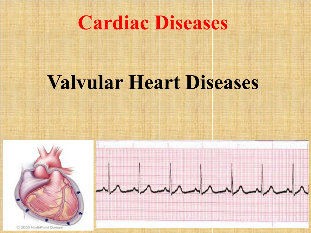
Cardiac Diseases Valvular Heart Diseases
Overview Rheumatic Heart Disease Mitral Stenosis (MS) Mitral Regurgitation (MR)
Rheumatic Heart Disease Rheumatic fever is an inflammatory disease that may develop after an infection with group of Streptococcus bacteria (such as strep throat or scarlet fever). The condition usually appears in children between the ages of 5 and 15, even though children and older adults. Rheumatic fever is a relatively serious illness that, if left untreated, can cause stroke, permanent damage to heart valves, and death. Usually results in distortion and scarring of the valves.
Mitral Stenosis (MS) Definition: It s a valvular heart disease characterized by the narrowing of the orifice of the mitral valve of the heart. As a result, less blood flows to the body. Back pressure which builds up behind the narrowed valve can cause various problems and symptoms. The more severe the narrowing, lead to more serious the problems. Normal Mitral Valve area: 4-6 cm2.
Etiology of Mitral Stenosis 1. Congenital factor. 2. Rheumatic fever is the predominant cause. 3. Rarely, other factors can cause mitral stenosis in adults. These include: a) Calcium deposits forming around the mitral valve. (Primary hyperparathyroidism and malignancy account for about 90% of cases of hypercalcaemia). b) Radiation treatment to the chest due to tumor.
MS Pathophysiology 1. Thickening and calcification of valvular tissue, thereby narrowing the MS opening and limiting blood flow from the left atrium to the left ventricle. Increased pressure in the left atrium, leading to pulmonary hypertension and left atrial hypertrophy. Right ventricular failure, producing from pulmonary congestion. 2. 3.
Possible clinical manifestations 1. Murmur is an early sign (as it for most of valve disorders). 2. Dyspnea on exertion (due to pulmonary venous hypertension) as the first symptom 3. Progressive fatigue (result of low cardiac output) 4. Right sided heart failure. 5. Peripheral edema 6. Orthopnea 7. Weak and often irregular pulse (because of atrial fibrillation) 8. Hemoptysis & cough due to pulmonary capillary pressure. 9. Repeated respiratory infections.
Possible diagnostic test findings: Chest X-ray: Enlargement of the left atrium and right ventricle and pulmonary congestion. Echocardiogram: Thickening of the mitral valve and left atrial enlargement. Electrocardiography (ECG): ST- segment depression and T-wave inversion. Angiography: Mitral Stenosis.
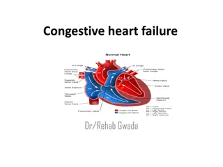
Possible medical complications:- 1. Atrial fibrilation (AF). 2. Thrombosis. 3. Congestive Heart Failure (CHF).
Treatment for Mitral Valve Stenosis 1. Beta blockers: decrease the force contractions of the heart muscles, and reduce blood vessel contraction e.g: Tenormin. 2. Calcium channel blockers: are prescription medications that relax blood vessels and increase the supply of blood and oxygen to the heart while also reducing the heart workload e.g: Amlodipine. 3. Anticoagulants (if showing signs of atrial fibrillation) e.g: Heparin. 4. Digoxin only for end-stage heart failure. 5. Diuretics.
Mitral valve stenosis surgery Deciding whether you need surgery and if so, when, depends on the severity of your disease, the surgery including:- Balloon Valvotomy: This procedure is the preferred treatment for mitral valve stenosis. A doctor uses a catheter and a tiny balloon to stretch open the narrowed valve. Repair surgery (Commissurotomy): This is typically an open-heart surgery using a heart-lung bypass machine. A surgeon removes calcium deposits and other scar tissue from the valve leaflets to widen the valve.
Mitral Regurgitation (Insufficiency)
Mitral Regurgitation Overview Mitral valve regurgitation is the most common form of heart valve disease. An estimated 4 million people in the U.S have significant mitral valve regurgitation. About one in 10 people age 75 and older have mitral valve regurgitation.
What is mitral regurgitation (MR)? Definition: Backflow of blood from the Left ventricle to the Left atrium during systole. OR incomplete closure of the mitral valve. When the two leaflets of the mitral valve do not close properly, mitral valve regurgitation occurs.
Mitral valve regurgitation causes: Following are some of the causes of mitral valve regurgitation: 1. Congenital defect. 2. Weakened heart muscle. 3. Mitral valve prolapsed. 4. Heart valve infection (Bacterial endocarditis). 5. Rheumatic fever.
MR Pathophysiology 1. Valvular incompetence. 2. To compensate , the left ventricle becomes hyperdynamic. In acute severe MR, the left atrial and pulmonary venous pressures increase quickly, leading to pulmonary congestion and pulmonary edema. 3. Increase left atrial pressure, pulmonary hypertension, and left atrial hypertrophy. 4. Left atrial enlargement predisposes the patient to atrial fibrillation and arterial thromboembolism. 5. In long-standing MR, patients may develop pulmonary hypertension which lead to right-sided heart failure.
MR Signs and Symptoms Acute severe MR, as occurs with papillary muscle rupture, is almost always symptomatic because the sudden regurgitant volume load in the non dilated left ventricle and atrium, leads to pulmonary venous hypertension and congestion. Patients with chronic MR may remain asymptomatic for years because the regurgitant volume load is well tolerated as a result of compensatory ventricular and atrial dilation.
MR Signs and Symptoms When symptoms do develop, the most common are: 1. Shortness of breath. 2. Cough. 3. Fatigue. 4. Orthopnea. 5. Peripheral edema. 6. Heart murmur. 7. Palpitations caused by atrial fibrillation.
Possible diagnostic test findings: 1. Chest X-ray may demonstrate left atrial enlargement and/or cardiomegaly. 2. Echocardiogram: Enlargement of the left atrium; abnormal movement of the mitral valve. 3. Angiography: Insufficiency in mitral valve. 4. Troponen test: Positive in acute MR.
Possible medical complication: 1. Embolism 2. Thrembosis 3. Congestive Heart Failure 4. Ruptured papillary muscle.
Mitral Valve Regurgitation - Surgery Deciding whether you need surgery and if so, when, depends on the severity of your disease, the surgery including:- Repair:- It s the preferred surgery instead of replacement of the valve, (reshape the valve by removing excess valve tissue). Replace:- With replacement, the damaged valve is removed and a mechanical (plastic or metal) or bioprosthetic valve (usually made from pig tissue) is stitched into place.
Nursing intervention for MS and MR 1. Keep the patient on a low-sodium diet. 2. Prepare the patient for valve replacement or percutaneous balloon valvuloplasty, as indicated. 3. Assess cardiovascular and respiratory status. 4. Keep the patient in semi-Fowler's position. 5. Monitor and record vital sign; Intake/ Out and ECG readings. 6.Teach the patient about diet restrictions. 7. Assess peripheral edema. 8. Give the patient oxygen therapy. 9. Give antibiotics as prescribed.


")
")


?")