Understanding Shock: Causes, Phases, and Pathophysiology
This comprehensive guide delves into the various aspects of shock, including hypovolemia, pulmonary embolism, and acute myocardial infarction. It covers the general concept of shock, circulatory failure, phases of shock, pathophysiology, manifestations, diagnostic approaches, and complications such as multiple organ dysfunction syndrome (MODS). The content explores the underlying mechanisms of shock, highlighting the importance of recognizing early signs and symptoms for prompt intervention.

Download Presentation

Please find below an Image/Link to download the presentation.
The content on the website is provided AS IS for your information and personal use only. It may not be sold, licensed, or shared on other websites without obtaining consent from the author. Download presentation by click this link. If you encounter any issues during the download, it is possible that the publisher has removed the file from their server.
E N D
Presentation Transcript
1. hypovolemia/shock 2. pulmonary embolism 3. acute myocardial infarction Ivan undrle, Pavel Suk, Jan Hruda ARK, FNUSA 2016
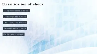
Shock Circulatory failure supply demand 2. 1. cardiogenic pump 2. obstructive obstruction 3. hypovolemic filling 4. distributive - shunts 1. 3. 4.
Phases of Shock 1. Compensation 2. Decompensation 3. Refractory Inflammatory cascade induction and organ damage - secondary-hit model Organ damage further increases inflammatory cascade induction vicious circle Each type of shock differs at the beginning, however during the late phase all types of shock look similar (like distributive shock)
Pathophysiology The main problem is cell hypoxia Stress response catecholamines, RAAS, cortisol, glucagon Systemic inflammatory response Imunity, inflamatory mediators Localy OK, but generalized response is harmful
Pathophysiology 1. Macrocirculation blood flow centralization rarely warm shock 2. Microcirculation Endothelium damage Increased vascular leakage, leucocytes adherence Main role in shock 3. Coagulation Intravascular coagulation 4. Metabolism Increased gluconeogenesis, proteolysis Lactate acidosis
MODS 1. Circulation Vasoplegia, cardiomyopathy 4. Coagulation DIC 5. CNS Altered consciousness 2. Lungs ARDS 6. GIT Loss of barrier function 3. Kidney AKI
Signs/Symptoms Nonspecific Variable Unreliable Tachypnea > 30 breaths/min, dyspnea Skin: Wet, cold CRT(> 2 s) Hypotension, tachycardia: SBP < 90 mmHg MAP < 60 mm Hg Tf > 100/min Cave compensatory shock/BB Mental state: Confusion Iritation coma Oliguria: diuresis < 0,5 ml/kg/hr for 1 6hrs
Diagnostics 1. Basic Lab BC, coagulation (Q/INR, aPTT, fib) ions, gly urea, kreatin CRP (sepsis?) 2. ABG Ventilation/oxygenation Lac, SvO2, ScvO2
ABG Lac ScvO2 Product of anaerobic glycolysis Non-toxic, serves also as a fuel normal < 2 mmol/l Mortality predictor Early sign O2ER = (SaO2 SvO2) / SaO2, normaly 25% normal SvO2 is 75% SvO2< 70% = O2supply impairment
Initial resuscitation Preload optimization increasing CO, fluids volume challenge , PLR Persistent hypotension catechols (norepinephrine) If CO does not rise with fluids, add inotropes (dobutamin) Lowering of inadequately high afterload (hypertension crisis)
Causal treatment 1. Cardiogenic shock: SCG - PCI Arrhythmia treatment (AV block III., VT) 2. Hypovolemic shock: fluids hemotherapy damage control surgery/damage control resuscitation 3. Obstructive shock: thrombolysis Pericardial effusion evacuation
Most common Causes of Hypovolemia Bleeding Loss of fluids (sweating, vomiting, diarrhea, ....) inadequate intake Burns 3rd space losses Ileus anafylaxis, sepsis (relativ hypovolemia)
Treatment 1. Initial resuscitation 2. Causal treatment Goal is to restore organ perfusion, O2 supply Early initiation Secondary goal: restoration of O2 transportation capacity (ERY...)
Venous access 2-3 thick peripheral cannulas Central venous access is secondary (good for catecholamine, not fluids) Exception: thick central lines (Edwars AVA 9F)
Arterial Catheter Continuous blood pressure monitoration accurate PPV Repeated blood draws
Witch fluid to use? Ions Na+ and K+ - ICT/ECT distribution Oncotic pressure plasma/ECT distribution 4% 4% ECT 20% ECT 20% ICT 40% ICT 40%
Glucose Inadequate Absolute water deficit Hypernatremia correction 4% 4% ECT 20% ECT 20% ICT 40% ICT 40% Distribution volume Distribution volume
Crystaloids Fast leak into the ECT compartment Substitution has to be 4x higher than the deficit (...recently questioned) swellings 4% 4% ECT 20% ECT 20% ICT 40% ICT 40% Distribution volume Distribution volume
Coloids Do not leave the intravascular compartment Equal the deficit Adverse reactions, contraindication sepsis renal damage Good for acute blood loss 4% 4% ECT 20% ECT 20% ICT 40% ICT 40% Distribution volume Distribution volume
Blood products Only for blood loss corrections 5% albumin natural colloid expensive
Fluid resuscitation goals Blood pressure, heart rate Centralization reversal diuresis Decrease of the PPV / SVV Sc(v)O2 a lactate normalization Filling pressures(CVP, PAOP) not a good target
Acute bleeding Blood loss fractures pelvis (5000ml) femur (2000ml) tibia (1000 ml) humerus (800 ml) radius (400ml) 15% (750 ml) well compensated 30% (1,5 l) tachycardia, oliguria, normotension however organ perfussion! More than 30%: hypotension, tachycardia, oligo-anuria, ...
Treatment Basic approach ... ABCD Stop the bleeding Give i.v. Fluids + catecholamine Blood type O- (4 immediately available), after 30 minutes type matched Fresh frozen plasma 1:1 with erythrocytes Target Hb 70-90 g/l, CNS trauma 100g/l Thrombocytes 50 100 tis/ul Fibrinogen 1,5 g/l Prevent hypothermia, hypotension and acidosis
AIM Myocardial ischemia Causes 1. 2. Increased demand tachycardia Low oxygen content anemia, CO poisoning, hypotension, pulmonary disease Low coronary artery blood flow 3. 90 % low coronary artery flow coronary atherosclerosis Transmural ischemia 3/4 of the myocardial wall (complete closure) Laminar/subendomyocardial 1/3 of the myocardial wall (partial closure + increased demand)
Diagnostics 1. Patinet history/clinical evaluation 2. ECG a Lab 3. ECHO, SCG STEMI Chest pain NSTEMI Chest pain AP History ECG Chest pain ST depression at least 1 mm and /or T wave inversion ST elevation at least 2 mm in leads V1 V3 or at least 1 mm in V4 V6, I, aVL, II, III, aVF. ST depression at least 1 mm and /or T wave inversion ST elevation in at least two adjacent leads. New LBBB or (RBBB + LAH, RBBB + LPH). Lab Positive TNT Positive TNT Negative TNT
Localization Anteroseptal V1-V4 Anterolateral V1-V6 Lateral I, aVL, V5, V6 Lower/diafragmatic II, III, aVF
Treatment Continuous vital signs / ECG IV access Oxygen 4 8 l/min 12 lead ECG Blood draw Lab /TNT Analgosedation - morphine ASA 500 mg i.v./200 400 mg p.o. heparin 5000 j i.v./enoxaparin 1 mg/kg s.c./i.v. clopidogrel 300 nebo 600 mg p.o. metoprolol i.v. If tachycardia
Cardiogenic Shock Severe, long-lasting arterial hypotension Low CO Increased filling pressure CVP/PAOP Alteration of consciousness, oliguria, cold periphery, sweat, cyanosis
Treatment Most important is to increase oxygen supply and lower oxygen consuption by myocardial muscle Preload optimalization: diuretics/fluids Afterload optimalization: vasodilatation / cave coronary arthery perfussion Inotropy dobutamin Treatment of the cause PCI/thrombolysis
Avoid Tachycardia short diastolic phase, increased work load (however, sometimes only chance how to increase CO) Severe hypotension, hypovolemia, vasodilatation low coronary artery perfusion pressure (Ao pressure EDP LV) Increased preload/afterload increase of wall tension, work
Treatment Oxygen increase O2 supply NIV, invasive ventilation oxygenation, decreases preload/afterload Diuretics/fluids decrease preload, in later phase optimization of preload (fluid challenge/PLR) Catecholamine norepinephrine for blood pressure, dobutamin (milrinon, levosimendan) for inotropy Vasodilatancia nitrates, coronary artery, but also systemic vasculature ( increased blood pooling, preload lowering; arterial afterload lowering) Morphine improves dyspnea
Pulmonary Embolism Sudden obstruction of pulmonary vasculature with emboli (blood cloth, fat, tumor, air/gas, foreign body, ...) Etiology: 85% low extremity/pelvic DVT
Risc Factors Virchov trias - venostasis, hypercoagulation, vessel wall damage Major surgery Lower extremity fractures Hypercoagulation (Leiden ...) Heart Failure (blood stasis) Sepsis (coagulation activation) High age (70 years) Immobilization Obesity Pregnancy Economy class syndrome corticoids, diuretics, HAC
Diagnosis History Sudden dyspnea, chest pain, tachypnea, cough, syncope, hemoptysis Clinical evaluation tachypnea, cyanosis, hypotension, shock, tachycardia, neck veins distension Lab ABG - hypoxia, hypocapnea, Ralc DD- negative practically excludes PE DD- positive tumors, inflammation, post-surgery, sepsis ...
Chest X-ray Excludes other reasons for dyspnea Fleischman sign- atelectasis Westerman sign decreased pulmonary vascularization
ECHO RV dilatation, paradoxical septum movements, pulmonary hypertension, Tri regurgitation
Other Vein US femoral, popliteal TEE thrombus in pulmonary artery Swan-Ganz - precapillary PH, high CVP, high RV pressure, increase PAP, Ventilation/perfusion scan low specificity
Managment Clinical probability, DD, echo and CT angio Clinical probability: Signs of DVT Other dg improbable 3 1,5 - - - low 0-1 (3,4%) moderate 2-6 (20%) high 7 (63%) Tachycardia 100 Immobilization more than 3 days, surgery within 4 weeks 1,5 1,5 - - 0-4 PE less probable More than 4 - PE highly probable DVT, PE in history hemoptysis malignancy 1,5 1 1
1. High risk PE (shock, hypotension) CT angio or ECHO, if CT unavailable/impermissible for the patient CT/ECHO positive - trombolysis
2.Low risk PE (without shock/hypotension) High clinical suspicion CT angio Low clinical suspicion DD Negative DD nearly completely exclude PE TNT, NT pro BNP, RV dysfunction thrombolysis/heparinization
Massive PE unstable or RV dysfunction, TNT, NTproBNP Thrombolysis optimally within 48 hrs alteplasis (0,9mg/kg) 10 mg bolus iv. + 90 mg cont. iv. for 2 hrs + heparin for min 72 hrs - UHF 80 IU/kg bolus + 18 IU/kg/hr


")