Overview of Intestinal Nematodes: Ascaris lumbricoides and Strongyloides stercoralis
Intestinal nematodes, including Ascaris lumbricoides and Strongyloides stercoralis, are common parasitic infections affecting the small intestine. Ascaris is a roundworm with a global distribution, transmitted via fecal-oral route or flies, while Strongyloides is a threadworm primarily found in tropical regions, requiring warm, moist soil for transmission. Clinical manifestations vary from asymptomatic to respiratory and gastrointestinal symptoms. Diagnosis involves identifying eggs or larvae in stool samples. Understanding their life cycles and clinical presentations is crucial for effective management.

Download Presentation

Please find below an Image/Link to download the presentation.
The content on the website is provided AS IS for your information and personal use only. It may not be sold, licensed, or shared on other websites without obtaining consent from the author. Download presentation by click this link. If you encounter any issues during the download, it is possible that the publisher has removed the file from their server.
E N D
Presentation Transcript
NEMATODES SMALL INTESTINE, SYSTEMIC NEMATODES
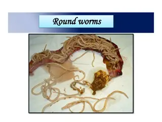
1- ASCARIS LUMBRICOIDES (ROUND WORM) Habitat: small intestine. Worldwide distribution mainly tropics. Transmission: fecal-oral; or by flies. Egg is very resistant, can survive years.
CLINICAL PICTURE AND DIAGNOSIS: Related to number of worms; small numbers are asymptomatic. Pulmonary phase: Ascaris pneumonitis. Intestinal phase: Large numbers of adults in intestine: colics, loss of appetite, diarrhea. Complications: intestinal obstruction, malnutrition, vitamin deficiency if in large numbers. Diagnosis: During migratory stage: eosinophilia, bronchial wash (may detect the larva), x-ray (cellular infiltration of the lung). During intestinal stage: stool examination for eggs.
2- STRONGYLOIDES STERCORALIS (THREADWORM) Habitat:small intestine. Mainly a tropical parasite because requires warm moist soil for transmission. The only important helminth that can complete its life cycle in the human host and increase its numbers. Facultative worm. Transmission: skin contact with invasive larvae (third stage filariform larvae) in soil, or by autoinfection.
LIFE CYCLE: Infective stage: third stage filariform larvae skin invasive. They mature to adults in submucosa of Small intestine. Small numbers of larvae get into blood vessels and circulate again to produce more adults (internal auto infective cycle) or invade perianal skin during their descend in the stool, and enter blood vessels to eventually produce new adults (external auto infective cycle).
CLINICAL PICTURE: Most asymptomatic. Cutaneous phase: itching and swelling occur at the site of penetration by the larvae. Pulmonary phase: Pneumonia due to migration of the larvae in the lung. Intestinal phase: by the adults present in the intestine, GI - peptic ulcer like symptoms, diarrhea, malabsorption which lead to steatorrhea (fatty stool). Hyperinfection:(disseminated immunocompromised; spread of larvae to peritoneum, lung, CNS with contamination of those organs with gram negative bacteria; transmural small intestine spread of larvae and bacteria with necrosis of intestine strongyloides) in
DIAGNOSIS: During migratory stage: eosinophilia, bronchial wash (may detect the larva), x-ray (cellular infiltration of the lung) During intestinal stage: Stool examination . Duodenal aspirate or Enterotest duodenal string test Serology (the most sensitive) Culture of stool (Harada-Mori or Baerman) allows "free living" strongyloides to multiply.
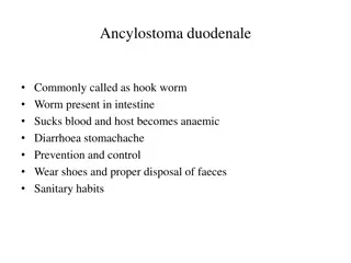
3- ANCYLOSTOMA DUODENALE (HOOKWORM): Habitat: small intestine. Transmission by contact of skin with soil contaminated with third stage filariform larvae. Infective stage: third stage filariform larvae.
LIFE CYCLE: Immature eggs in feces hatch to infective (filariform) larvae in 7 days larvae penetrate skin of host (e.g. bare feet). As adults, they attach by mouth to small intestinal mucosa and suck blood.
CLINICAL PICTURE: 90% asymptomatic. Cutaneous phase: itching and swelling occur at the site of penetration by the larvae. Pulmonary phase: Pneumonia due to migration of the larvae in the lung. Intestinal phase: by the adults present in the intestine. Attachement of the worms to the mucosa with their buccal capsules and teeth. They eat the tissues and suck the blood. Manifested as abdominal pain, polyphagia, reddish stool. Heavy infections (20 - 100 worms) Iron deficiency anemia (IDA) Malnutrition from protein loss (hypoproteinaemia)
DIAGNOSIS: During migratory stage: eosinophilia, bronchial wash (may detect the larva), x-ray (cellular infiltration of the lung). During intestinal phase: stool examination for ova. Sometimes, egg counting occurs by stoll technique or kato technique in order to diagnose intensity of the infection.
SYSTEMIC NEMATODES: 1-TRICHINELLA SPIRALIS: This is a zoonosis infecting most carnivorous mammals; especially pigs. Man infected by eating Trichinella infected uncooked meat. Common in geographic areas where undercooked pork is eaten; 5-15% of North American population infected at some time.
LIFE CYCLE: Encysted larvae in meat, when eaten, excyst (hatch) and penetrate into small intestine submucosa: where they mature to adults in 1-2 weeks: producing larvae which penetrate blood vessels and disseminate to all muscles. There, they cause inflammation and encyst in muscle cells (not cardiac muscles), remaining viable and quiet for many years.
CLINICAL PICTURE AND DIAGNOSIS: Clinical: Early (1-2 weeks): abdominal pain, diarrhea. Midterm (2-6 weeks): myalgia, muscle weakness, facial edema, rash; sometimes encephalitis and myocarditis. Long term (months): usually asymptomatic despite presence of trichinella "cysts . Diagnosis: Clinical picture with laboratory support (eosinophilia and raised creatine phosphokinase (CK). Microscopic examination of muscle biopsy. Serology.
2- TOXOCARA CANIS This is a zoonotic roundworm with the dog as reservoir. Uncommon human infection but consequences serious. Transmission is dog fecal (dog)-oral (human). Dog feces especially in sand and parks where children play. Eggs in soil remain viable and infective for several months.
LIFE CYCLE: Adult has cycle in dog the same as Ascaris in man. Man is an accidental "dead end" host. Eggs ingested by man/child, hatch after stomach passage and larvae migrate through small intestinal wall into vasculature and then to liver and lungs and beyond. Do not mature to adults but cause local inflammation especially in liver.
CLINICAL PICTURE AND DIAGNOSIS: Clinical: Hepatomegaly, pneumonitis, encephalitis, fever and eosinophilia in heavy infections. Retinal lesion : focal retinitis when single larva reaches retina. Diagnosis: Clinical syndrome with very high eosinophilia. Serology. Nothing in stools as man is an end host.
3- DRACUNCULUS MEDINESIS: Habitat: in the connective tissues. Exists usually in dry climate, where people rely on wells for their water supply. Present in Yemen, Sudan, Saudi Arabia, Nile valley, India and Ethiopia. It is the largest nematode. Man become infected by drinking of unfiltered water. Infective stage: L3 larvae.
CLINICAL PICTURE AND DIAGNOSIS: Clinical: 1-During migration of the gravid female, allergic manifectations appear in the form of nausea, dizziness and oedema. 2-Once reaching the skin, blister appears in the skin. 3-On rupturing of the blister, the worm protrudes from a tiny hole. Diagnosis: Place water on the blister present in the skin, rupture of the blister occurs within few minutes, the worm is protruded. Collection of this water, centrifuge, put on a slide, larvae could be seen.
4- WUCHERERIA BANCROFTI Habitat: Adults live in afferent lymphatic vessels (mainly of the lower half of the body). Microfilariae circulate the blood from 10:00 p.m. to 2 a.m. - this corresponds to peak activity of vector mosquitoes (culex). Infective stage: 3rd stage filariform larvae formed in culex. Maturation to adult requires several months.
CLINICAL PICTURE: Asymptomatic microfilaremia. Acute stage: Lymphangitis and lymphadenitis. Presented by fever, pain and redness over the affected limb. Orchitis and epididymitis. Chronic stage: 1-Elephantiasis (enlargement of one or more of limbs, srotum, breast or vulva). Its mechanism is: The inflammation ends in fibrosis, obstruction of lymphatics and leakage of fluid rich in protein under the skin. Presented by hard oedema. 2-Chyluria (passage of chyle with the urine) due to rupture of the obstructed lymphatics to the urinary passages.
DIAGNOSIS: Blood examinations (esp. Night blood) for microfilaria. Knott s technique is used if the number of microfilaria is scanty in the blood. Serology (IFAT, ELISA). Antigen capture (dip stick test). Quantitative buffy coat, to detect the DNA of the parasite, giving fluorescence. Urine examination to detect microfilaria in case of chyluria.

")


")


:")