Intestinal Helminths: Classification, Nematodes, and Common Infections
Intestinal helminths, including nematodes, are classified into unicellular and multicellular parasites. Nematodes are elongated worms found in the human body, causing common infections such as Enterobius vermicularis. The pathology of Enterobius infections often presents with pruritus ani. Learn about the types and characteristics of these parasitic organisms.

Uploaded on Sep 26, 2024 | 0 Views
Download Presentation

Please find below an Image/Link to download the presentation.
The content on the website is provided AS IS for your information and personal use only. It may not be sold, licensed, or shared on other websites without obtaining consent from the author. Download presentation by click this link. If you encounter any issues during the download, it is possible that the publisher has removed the file from their server.
E N D
Presentation Transcript
Intestinal Helminths Dr. Ibrahim Alkhalife
CLASSIFICATION OF PARASITES PROTOZOA HELMINTHS Unicellular Single cell for all functions 1: Amoebae: move by pseudopodia 2: Flagellates: move by flagella 3: Ciliates: move by cilia 4: Apicomplexa (Sporozoa) tissue parasites Multicellular Specialized cells Round worms (Nematodes): - elongated, cylindrical, unsegmented. Flat worms: - Trematodes: leaf-like, unsegmented. - Cestodes: tape-like, segmented.
Nematodes: General features Elongated worm, cylindrical, unsegmented and tapering at both ends. Variable in size, measure <1 cm to about 100cm. Sex separate and male is smaller than female 1. 2. 3.
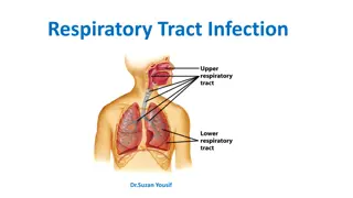
Nematodes: Location in the human body Intestinal nematodes Tissue nematodes
Nematodes: common intestinal infections Enterobius (Oxyuris) vermicularis (Pinworm, seatworm, 1. threadworm) Trichuris trichiura (whipworm) 2. Ascaris lumbricoides (roundworm) 3. Ancylostoma duodenale & Necator americanus 4. (hookworms) Strongyloides stercoralis 5.
1- Enterobius vermicularis (THREAD WORM) (Common names: Pin worm, seat worm( Found all over the world but more common in temperate regions. Children are more often evolved than adults, it tends to occur in groups living together such as families, army camps or nursery. Adult worms are mainly located in lumen of cecum and the female migrate to rectum to deposit her eggs on perianal skin.
Direct human - human infection occurs mainly by swallowing the eggs. Autoinfection occurs by contamination of the fingers. It can be seen by naked eye as white thread 1cm. Male is smaller than female 0.5cm, with coiled end.
Enterobius vermicularis (Oxyuris) Pathology Most infections are asymptomatic Main clinical presentation pruritus ani which can be very troublesome and occurs more often during the night, persistent itching may lead to inflammation and secondary bacterial infection of the peri-anal region
Enterobius vermicularis (Oxyuris) Infected children may suffer from emotional disturbance, insomnia, anorexia, loss of weight and loss of concentration and enuresis. Ectopic enterobiasis occurs in infected adult female when invade vulva and vagina result in valvo-vagintis, salpingitis, also adult worm can lodge in the lumen of appendix cause appendicitis.
Enterobius vermicularis (Oxyuris) DIAGNOSIS : Unlike other intestinal Nematodes, the eggs are not usually found in feces. The best method is to look for them around the anus by taking an anal swab or by using CELLULOSE ADHESIVE TAPE the examination should be done before defecation or bathing. Treatment Albandazole or Mebendazole for whole family
Enterobius vermicularis (Oxyuris)
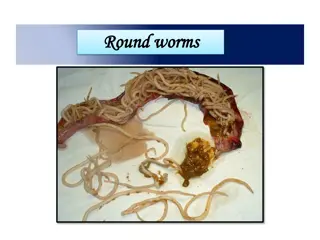
Ascaris lumbricoides (roundworm)
Ascaris lumbricoides (roundworm) The commonest human helminths infection all over the world. The large round worm which is normally located in the small intestine Found in jejunum and upper part of ileum Female 20 cm longer than male 10 cm Feed on semi digested food.
Ascaris lumbricoides (roundworm) Infective stage: embryonated egg Diagnostic stage: unembryonated egg
Ascaris eggs Ascaris larva emerging from egg Ascaris egg (embryonated)
Ascaris lumbricoides (roundworm) Pathology: 1-Adult worm: (small intestine) Light infection Heavy infection : intestinal obstruction Migrating adult : to bile duct-jaundice 2-Larvae: Loeffler`s syndrome Pneumonitis and broncho-spasm, cough with bloody sputum, Eosinophilia, urticaria : asymptomatic
Ascaris lumbricoides (roundworm) Loeffler`s syndrome: Larvae in lung pneumonia, cough, bloody sputum
Ascaris lumbricoides (roundworm) Ascaris larva in lung
Ascaris lumbricoides (roundworm) Diagnosis: -eggs in stool. -larvae in sputum. -adult may pass with stool. Treatment: Albendazole or Mebendazole
Trichuris trichiura Infective stage: embryonated egg Diagnostic stage: egg in stool
Trichuris trichiura (whipworm) World wide, common in poor sanitation It coexists with Ascaris because of similar requirements (the eggs needs 3 weeks in the soil to be embryonated which is the infective stage). Adult live in large intestine especially caecum and appendix in heavy infection the whole length of large intestine affected. Male and female worm have narrow anterior portion penetrate the intestinal mucosa
Trichuris trichiura (Whipworm) Pathology light infection: asymptomatic heavy infection: abdominal pain, bloody diarrhea. Rectal prolapse in children is a common complication.
Trichuris trichiura (Whipworm) Diagnosis: egg in stool characterized by its barrel shape with mucoid plugs at each pole. Treatment: Albendazole.
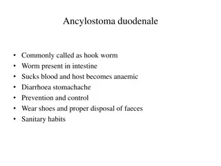
Hook worms Ancylostoma dudenale & Necator americanus Its buccal capsule (mouth) lined with hard hooks, triangular cutting plates and anticoagulant glands.
There are no specific symptoms or signs of hookworm infection but they give rise to a combination of: Intestinal inflammation Progressive iron-deficiency anemia & Protein deficiency
Filariform Larval (infective stage) invasion of the skin can produce a skin disease called: cutaneous larva migrans (creeping eruption) this is commonly caused by walking barefoot through areas contaminated with fecal matter. Larva migrate through the vascular system to the lungs, and from there up the trachea, and are swallowed. They then pass down the esophagus and enter the digestive system, finishing their journey in the small intestine where the larvae mature into adult worms. They mate inside the host, females laying up to 30,000 eggs per day, which pass out in feces (diagnostic stage). The eggs need to be in soil for about one week to become FILARIFORM LARVA
Hook worms Pathology& clinical picture - larvae: At the site of entry of larvae intense itching (ground itch) and dermatitis. Migration phase cough with bloody sputum pneumonitis and bronchitis but less sever than Ascaris, eosinophilia urticaria. - Adult worm: low worm burden (INFECTION): no symptoms. Moderate to heavy burden: Epigastric pain, vomiting, hemorrhagic enteritis. Protein loss: hypo-proteinaemia edema. Anemia: due to withdrawal of blood by parasites and hemorrhage from punctured sites lead to sever anemia = microcytic hypochromic anemia.
Hook worms Diagnosis and treatment Diagnosis: -Eggs in stools; -occult blood (+) Treatment: Albendazol, Mebendazole
Strongyloides stercoralis Widely distributed in tropical area at Asia, Africa & South America Fatal dissemination in immuno-compromised host. It is smallest pathogenic nematodes 2.5mm. adult live membrane of duodenum, jejunum rarely mucous membrane of bronchus Autoinfection is very important criteria
Strongyloides stercoralis life cycle The parasite shows 3 different modes of development: 1-Direct development: The rhabditiform larva pass from stool and become directly a Filariform larva if the environment of the soil is suitable. 2-Indirect development: In external environment Rh. larva becomes free living adults, produce eggs, rhabditiform larva and Filariform larva (Infective stage). 3-AUTOINFECTION: Internal: when the rhabditiform larva become a filariform larva in the intestine and penetrate the intestine External: fecal contamination of skin Rh larva > filariform penetrates the skin
Strongyloides stercoralis LIFE CYCLE
Strongyloides stercoralis Pathology and clinical picture Cuteneous little reaction on penetration. sever dermatitis at perianal region in case of external autoinfection Migration: pneumonitis during larval migration. Intestinal: inflammation of upper intestinal mucosa, diarrhea, upper abdominal pain in the epigastria colicky in nature. Disseminated strongyloidiasis: in patient with immunodeficiency, uncontrolled diarrhea granulomatus changes necrosis--perforation--peritonitis death.
Strongyloides stercoralis Diagnosis rhabditiform larvae diagnostic stage in: -Stool examination -Duodenal aspirate Treatment Albendazole, Mebendazole
Common Tapeworm Infections TAPEWORM LAB. CLINICA L PICTURE DIAGNOSI S TRANSMISSION OF INFECTION LOCATIO N OF ADULT IN HUMANS Small Intestine LOCATION OF LARVA IN HUMANS DISEASE eggs or proglottids in stools Taenia saginata taeniasis ingestion of larva in undercooked beef not present vague digestive disturbances vague digestive disturbances Taenia solium- Small Intestine eggs or proglottids in stools taeniasis ingestion of larva in undercooked pork not present ADULT depending on locality: from none to epilepsy X-ray, CT, MRI Serology Taenia solium- LARVA (cysticercus cellulosae) not present (except in Autoinfection, small intestine) sub- cutaneous muscles brain, eyes ingestion of egg Cysticercosis hymenolepiais ingestion of egg Small Intestine Intestinal Villi eggs in stools Hymenolepis nana Enteritis diarrhoe a depending on locality X-ray, CT, US Serology Hydatid sand ingestion of egg Echinochocc us granulosus hydatid disease not present Liver, lungs, Bones etc
Taenia saginata Is an obligatory parasite of man, the adult worm live in the SMALL INTESTINE CATTLE become infected by ingesting grass contaminated with eggs or gravid segments which passed from human faeces. In the cattle the onchosphere hatches out go to circulation and transformed to cysticercus stage in the muscle known as CYSTICERCUS BOVIS Man become infected by eating undercooked or improperly cooked beef, the adult worm lives in small intestine of man passing eggs and gravid proglottids to the environment. The majority of cases are Asymptomatic, some patients have vague intestinal discomfort, vomiting and diarrhoea
Hymenolepis nana
Location of hydatid cyst Echinococcus granulosus
Hydatid cyst


")


")


")
")

")
")
")


 invasion of the skin can")