Parasitic Worms: Nematodes and Cestodes Overview
Explore the world of parasitic worms with a focus on Nematodes and Cestodes. Learn about Wuchereria bancrofti causing lymphatic filariasis and Echinococcus species leading to hydatid disease. Discover the transmission, laboratory diagnosis, treatment, and clinical aspects associated with these pathogenic worms.

Download Presentation

Please find below an Image/Link to download the presentation.
The content on the website is provided AS IS for your information and personal use only. It may not be sold, licensed, or shared on other websites without obtaining consent from the author. Download presentation by click this link. If you encounter any issues during the download, it is possible that the publisher has removed the file from their server.
E N D
Presentation Transcript
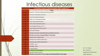
Nematodes & Cestodes Lab-6-
Nematode(round worm) Wuchereria bancrofti Wuchereria is thread-like worm that lies coiled in the lymphatic vessels. Wuchereria bancrofti causes lymphatic filariasis ( Elephantiasis). Transmission and reservoirs: W. bancrofti, transmitted primarily by mosquitoes of the Anopheles or Culex species. Infective stage: larva 3
Wuchereria bancrofti Laboratory Diagnosis: Thick blood smears taken from the patient at night reveal the microfilariae. Treatment: Diethylcarbamazine citrate (DEC)is the drug of choice
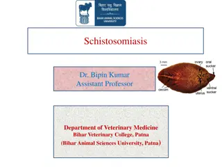
Cestodes (Tape worm) Echinococcus Echinococcus multilocularis (Hydatid worm or Hyper Tape-worm). The larva of Echinococcus tapeworm) causes unilocular hydatid cyst disease. Multilocular hydatid Echinococcus multilocularis, which is a minor pathogen. E. granulosus is composed undifferentiated neck proglottids, making it tapeworms granulosus and Echinococcus granulosus (dog disease is caused by of a scolex, three smallest region one and of only the
Echinococcus Transmission: Dogs, the definitive host, are infected by eating contaminated meat, humans like the usual intermediate hosts, become infected following ingestion of eggs passed in dogs feces. Clinical disease: In humans, E. granulosus cause echinococcosis (hydatid disease). A. Most cyst occur in the liver or lungs. B. Cysts in bone, the CNS, heart, and kidney carry a serious prognosis.
Echinococcus Diagnosis: 1. Radiographs, ultrasound, and computed tomography (CT) scan are helpful visualizing cysts. 2. Routine histology: Echinococcus species can detected in aspirates or biopsies of liver or lungs. 3. Serological tests. Treatment: Albendazole is the drug of choice. Surgical removal of cysts may follow successful treatment.

")
")


")