Overview of Malignant Catarrhal Fever in Cattle and Other Species
Malignant catarrhal fever (MCF) is a fatal disease affecting various species, caused by two different viruses transmitted from wild animals. It is characterized by low morbidity but high mortality rates, with distinct clinical signs such as sudden death, head and eye manifestations, and intestinal issues. The disease has specific transmission modes and commonly impacts cattle, bison, elk, sheep, and goats. Early detection and preventive measures are crucial in managing MCF outbreaks.

Download Presentation

Please find below an Image/Link to download the presentation.
The content on the website is provided AS IS for your information and personal use only. It may not be sold, licensed, or shared on other websites without obtaining consent from the author. Download presentation by click this link. If you encounter any issues during the download, it is possible that the publisher has removed the file from their server.
E N D
Presentation Transcript
MALIGNANT CATARRHAL FEVER Dr Padma Nibash Panigrahi Assistant Professor Department of Veterinary Medicine DUVASU, Mathura
MALIGNANT CATARRHAL FEVER (BOVINE MALIGNANT CATARRH, MALIGNANT HEAD CATARRH Etiology : Really two diseases (clinically and pathologically indistinguishable) associated with two different infectious agents with different ecologies 1. Alcelaphine herpesvirus-1 (AHV-l) wildebeest-associated MCF virus, transmitted to cattle from blue wildebeest 2. Ovine herpesvirus- 2 (OvHV-2) sheep-associated MCF virus transmitted to cattle from sheep Neither agent transmit from cattle to cattle neither of the viruses cause any disease in their principal host, the wildebeest and the sheep
Geographic Distribution AHV-1 primarily in Africa Carried by wildebeest, antelope Also in zoological and wild animal parks OHV-2 worldwide Carried by domestic and wild sheep and goats Major cause of MCF worldwide
Species Affected Susceptible species Cattle, bison, elk, reindeer, moose, domestic pigs, giraffe, antelope, red and white-tailed deer, nilgai Carrier species Sheep, goats, wildebeest, antlope Morbidity- low Mortality- high (100%)
Transmission AHV-1 Inutero Contact with nasal and ocular secretions Aerosols during close contact OHV-2 Respiratory (aerosol) Transplacental rare Contact with nasal secretions
Clinical Signs 1. Peracute form: sudden death 2. Head and eye form Majority of cattle cases 3. Intestinal form Initially like head and eye form, but death occurs from severe diarrhea 4. Mild form Inoculated animals; recovery expected
Head and Eye Form: Early Stages Reddened eyelids Bilateral corneal opacity Crusty muzzle, nares Nasal discharge Salivation
Head and Eye Form: Later Stages Erosions on the tongue, buccal mucosa, necrosis, ulcer in oral cavity Erosions on the buccal mucosa
Clinical Signs in Bovidae Swelling of Joints, superficial lymph nodes Sloughing of Horn, hoof coverings Nervous signs Incoordination, head pressing, nystagmus, hyperesthesia Swollen pre-scapular lymph node
Peracute and alimentary tract forms short course of 1-3 d high fever dyspnoea acute gastroenteritis resembles the 'head and eye' form except that there is marked diarrhoea Mild form transient fever mild erosions -oral and nasal mucosa. may be followed by complete recovery
Post Mortem Lesions Erosions on the tongue and soft and hard palate
Post Mortem Lesions Necrotic areas in the omasal epithelium Multiple erosions of intestinal epithelium
Post Mortem Lesions Greatly enlarged lymph node compared to normal Necrotic areas in the larynx Diptheritic membrane often present
Post Mortem Lesions Urinary bladder mucosa hyperemic and edematous Kidney often has raised white foci on the cortex
Collection of Samples for diagnosis Histology Formaline fixed brain, lymph node, alimentary tract mucosa, pharynx, oesophagus, rumen, liver, adrenal gland, kidney, urinary bladder, salivary gland Virology lymph node, spleen, lung (PCR)
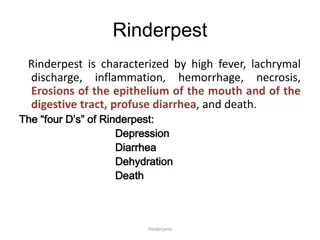
Differential Diagnosis Salmonellosis Pneumonia complex Oral exposure to caustic materials Mycotoxins Poisonous plants BVD mucosal disease Bluetongue Rinderpest FMD Vesicular stomatitis
Laboratory Diagnosis Histopathology PCR Virus isolation (AHV-1)- Madin Darby bovine kidney cell line (MDBK) Serology AHV-1 antibodies in wildebeest Immunofluorescence, immunoblot, VN, ELISA, immunocytochemistry OHV-2 antibodies in sheep Immunofluorescence, immunoblot
Treatment Survival is rare if clinically ill Mortality reaches 100% No specific antiviral drug Antibiotics for secondary bacterial infection Supportive therapy Recovered animals will remain virus carriers
Control Isolation of affected animal Separation of cattle from sheep in outbreak (sheep transmit diseases) Wild life 1 KM distance from livestock Quarantine Hygiene Vaccination ??? ( no specific immunity)