Understanding Spiruridae and Filariidae Nematodes in Vertebrates
Spiruridae and Filariidae are families of parasitic nematode worms affecting vertebrates and insects. This article delves into the characteristics, hosts, geographical distribution, biology, and control measures of Spiruridae, focusing on Spirura rytipleurites, and Filariidae, highlighting Dirofilaria immitis and diseases caused by these parasites.
Download Presentation

Please find below an Image/Link to download the presentation.
The content on the website is provided AS IS for your information and personal use only. It may not be sold, licensed, or shared on other websites without obtaining consent from the author. Download presentation by click this link. If you encounter any issues during the download, it is possible that the publisher has removed the file from their server.
E N D
Presentation Transcript
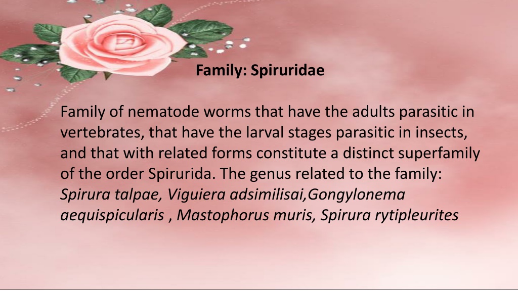
Family: Spiruridae Family of nematode worms that have the adults parasitic in vertebrates, that have the larval stages parasitic in insects, and that with related forms constitute a distinct superfamily of the order Spirurida. The genus related to the family: Spirura talpae, Viguiera adsimilisai,Gongylonema aequispicularis , Mastophorus muris, Spirura rytipleurites
Spirura rytipleurites (Filaria gastrophila) Introduction: Spirura from the name of the group (round) and ryti = wrinkled cuticle and pleurites for the location of the cysts in the leural cavity of the intermediate host. These worms were originally described from larvae recovered from the body cavity of the oriental cockroach or beetles and sometimes frogs (intermediate host).while the cat and accidentally sometimes the rat, Mongoose, Fox, hedgehog, and striped polecat regarded as definitive hosts. Geographical distribution: These worms are common in North Africa and probably into parts of southern Europe. Reported on infections in cats, one from Lyon and one from Madagascar. Also reported from a cat in Egypt. Site of infection: The adults live in the wall of the esophagus and stomach. Biology: The adults of this genus are recognized by the presence of one or two ventral cuticular bosses (inflations) in the cervical region. The eggs are ovoid with a smooth thick shell, and contain a first-stage larvae with a cephalic hook when passed in the feces.
The larvae were capable of developing in cockroaches that become infected when they ingest the eggs. The larvae grow within capsules in the abdominal cavities of the cockroaches where they become quite long, over a cm in length. The third stage larvae within the abdominal cavity of the insects have well developed reproductive systems. Cats become infected through the ingestion of infected cockroaches. Diagnosis: Can described and illustrated microscopy from specimens collected Pathogenesis: The nematode was identified as belonging to the genus Spirura. The number of parasites in this animal was considered significant and contributed to the death of the animal. Control/Prevention: As with many of the parasites, it is important to keep cats from hunting. At the same time, the large numbers of oriental cockroaches that can be present in houses would mean that in many parts of the world, infections could be very hard to control. by light and scanning of electron hosts. from the stomach final
Family: Filariidae The family causes diseases with economic importance by parasitic worms that are transmitted by the bite of blood-feeding insects. This debilitating group of diseases includes river blindness and lymphatic filariasis (commonly known as elephantiasis) in human as well as to animals (granulomatous nodule, pulmonary dirofilariasis).
Hosts: Dogs, foxes, wolves, coyotes, and cats. Dirofilaria immitis (Dog heartworms) Introduction: Dirofilaria immitis is a parasitic nematode responsible of canine and feline cardiopulmonary dirofilariasis in both domestic and wild hosts, and the causal agent of human pulmonary dirofilariasis. It is a zoonotic parasitic disease mainly located in temperate, tropical, and subtropical areas of the world.
Transmission: Dirofilariasis is transmitted neither person-to-person nor personto-mosquito-to- person. The transmission of dirofilariasis requires mosquitoes as the intermediate host as well as the production of microfilariae, which does not take place in humans. Different species of mosquitoes (Culex spp., Aedes spp., Anopheles spp.) act as an intermediate stage in order to complete their life cycle. Heartworm infection is a severe and life-threatening disease. Initially the pulmonary vasculature is affected, and the lung itself and, finally, the right chambers of the heart. However, the development of the parasite in cats takes longer compared to dogs and most infections are amicrofilaraemic. Additionally, many cats tolerate the infection without any noticeable clinical signs or with signs manifested only transiently and sometimes sudden death may arise without warning. Geographical distribution: Dirofilaria immitis is cosmopolitan in dogs particularly prevalent in warmer areas. Biology: Dirofilaria immitis has an indirect life cycle. The adult parasite sexually reproduces in its vertebrate host, and the larvae are transferred to the intermediate host, which is usually a mosquito or a flea
The life cycle described as following: 1. The life cycle starts with introduction of L3 larvae from an infected mosquito into the canid host. 2. In the dog, the larvae mature into L4 larvae and then into adult worms that localize in the pulmonary arteries and occasionally in the right cardiac ventricle. 3. Following a prepatent period of 6 to 7 months, microfilariae are produced via sexual reproduction and are released into the dog s bloodstream, where they may remain viable for months to years. 4. Mosquitoes feeding on the infected host ingest the microfilaria, which then migrate to the Malpighian tubules where they mature into first-stage larvae. 5. Ultimately, the larvae mature to the L3 stage in the salivary glands of the mosquito and become infective, thus completing the heartworm life cycle.
Pathogenesis: respiratory signs and acute death. In cases of pulmonary larval dirofilariasis, infected cats can have a Heartworm Associated Respiratory Disease (HARD), that is associated with asthma-like clinical signs.Cats are inherently resistant to heartworm infection. This is reflected by relatively low adult worm burden, the lack of microfilaraemia and their short life span, which complicates the diagnosis of this disease in the feline patient. Diagnosis: Dirofilariasis is diagnosed by detection of the microfilariae in the peripheral blood, immunological tests and molecular approaches also can be use. Dirofilaria immitis is usually diagnosed by the finding of the distinctive coin lesions on chest X-rays. The species that produce subcutaneous nodules are diagnosed by the finding of adult worms in biopsy specimens of these nodules. the infection of cats occurs asymptomatically or chronic
Treatment: surgical removal of lung granulomas and nodules under the skin; this treatment is also curative. In many cases, no treatment with medicines is necessary. Control and prevention: Dirofilariasis can be prevented by avoiding mosquito bites in areas where mosquitoes may be infected with Dirofilaria larvae. The risk of such mosquito bites can be reduced by leaving as little skin exposed as possible, by the use of insect repellent when exposed to mosquitoes, and by sleeping under an insecticide-treated bed net in areas where Dirofilaria-infected mosquitoes bite at night and The definitive treatment of Dirofilaria infection in humans is have access to sleeping areas.
Dirofilariasis in Human Humans become infected with Dirofilaria through mosquito bites. In persons infected with D. immitis, dying worms in pulmonary artery branches can produce granulomas (small nodules formed by an inflammatory reaction), a condition called pulmonary dirofilariasis. The granulomas appear as coin lesions (small, round abnormalities) on x-rays. Most persons with pulmonary dirofilariasis have no symptoms. People with symptoms may experience cough (including coughing up blood), chest pain, fever, and pleural effusion (excess fluid between the tissues that line the lungs and the chest cavity). Coin lesions on chest x-rays are not diagnostically specific for pulmonary dirofilariasis. Therefore, discovery of these lesions has led to invasive diagnostic procedures to exclude other, more serious causes, including cancer. Rarely, D. immitis worms have been found in humans in brain, eye, and testicle
Humans acquire the infection in the same manner as dogs, by the bite of a mosquito, but it is probable that most of the infective larvae die shortly after, with the infection resolving unrecognized and without causing any specific symptom. No predisposing factors are known to explain why in some cases larvae may develop further. After the bite of an infective mosquito, a stronger reaction with erythema, swelling and pruritus lasting 5 8 days. In most of the cases a single worm develops, probably because the stimulation the development of others. In rare cases the worm may develop to a mature adult and even fertilized worms releasing microfilariae have been described, patients, which in very rare cases may even reach the bloodstream. Dirofilaria repens: the developing stages of D. repens migrate subcutaneously for weeks up to several months in several parts of the body, usually with mild and unrecognized symptoms and sometimes causing irritation and erythema due to larva migration. The larva may reach the eyes and becoming visible through the subconjunctiva. Local surgery can be whitish worm from the site of infection. of the immune system prevents especially in immunosuppressed performed to remove a long
Other sites of infection: After weeks to several months from the infection, D. repens may stop to migrate and form a nodule of about one centimeter. In most cases, the nodules develop subcutaneously. various human body areas and tissues, mostly in the superficial tissues of the facial regions, as perioral and periorbital tissues, forehead, skin of the lower leg, soft tissues of the hand or finger, subcutaneous tissue of the hypogastrium and of the neck. Scrotum and testicles are other predilection site infested with larvae and, to a lesser extent, the breasts of women. Various reasons have been hypothesized for these preferences, such as lower body awareness of patients for these body parts or a tropism of D. repens due to abundance concentrations of sexual hormones. The nematodes could reach lymph nodes, the abdominal cavity, muscles and even the dura. However, D. repens if left untreated may survive Nodules have been reported in temperature of these areas, higher for up to one year.
Human pulmonary and dirofilariasis (D. immitis) Eyes and subcutaneous tissues of human infected with D. repens
