Diagnostic Modalities in Spine: An Overview
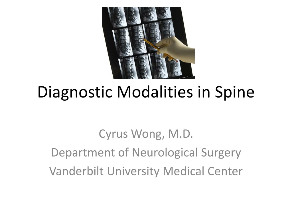
This detailed overview discusses various diagnostic modalities used in spine evaluations, including plain X-rays, X-ray with localization, CT scans, and MRI. Each modality's benefits and applications are explored, aiding in understanding different imaging techniques for spine pathology assessments.

Download Presentation

Please find below an Image/Link to download the presentation.
The content on the website is provided AS IS for your information and personal use only. It may not be sold, licensed, or shared on other websites without obtaining consent from the author. Download presentation by click this link. If you encounter any issues during the download, it is possible that the publisher has removed the file from their server.
E N D
Presentation Transcript
Diagnostic Modalities in Spine Cyrus Wong, M.D. Department of Neurological Surgery Vanderbilt University Medical Center
Plain Xray Historically used to diagnose all types of pathology related to the spine It relies on ionizing radiation to provide images The more dense an object the brighter the image That is why bone appears white on plain x-rays
Xray Cont. Used to help with localization in the operating room Allows to evaluate alignment Provides information with regards to stability Provides information about hardware placement
Cervical Spine Xray AP LAT
Thoracic Spine Xray Lat AP
Lumbar Spine Xray AP LAT
CT Scans A computed tomography (CT) scan uses x-rays to make detailed pictures of the spine It has the ability to reconstitute the images in the axial, sagittal and coronal plane It gives higher resolution pictures when compared to plain x-ray Excellent modality to evaluate fractures (spine trauma) Gives more information about soft tissue when compared to plan x-ray but not as sensitive as MRI Less time consuming than MRI
Cervical Spine Sagittal CT Axial CT showing fracture
C-Spine Cont. Coronal Sagittal
Lumbar and Thoracic CT Axial Sagittal
Thoracolumbar Fractures Compression Fracture Fracture Disslocation
MRI Uses a magnetic field and pulses of radio wave energy to make images Excellent image quality for soft tissue when compared to plain xray and CT The study of choice to evaluate soft tissue such as intervertebral disk, thecal sac, ligaments, joints and spinal cord Contrasted study is widely used in the detection of tumors
MRI Cont. Various sequences give different information T1 sequence Anatomic image Fat (bone Marrow) and Subacute blood (>48 hrs. old) appear bright (white) CSF and bone appear dark Most pathology appears dark but can enhance with contrast
MRI Cont. T2 Sequence Used to evaluate pathology Most pathology appears bright (white) Edema and CSF appear bright Bone and Fat appear dark Healthy Intervertebral disk appear bright
C-Spine MRI Sagittal T1 Axial T2 Sagittal T2
Cervical Stenosis Axial T2 Sagittal T2
Lumbar MRI Sagittal T2 Axial T2
Herniated Lumbar Disc Sagittal T2 Axial T2
EMG and Nerve Conduction Studies Studies used to help localize the site of compression Help delineate radiculopathy from neuropathy, myopathy or myelopathy Highly operator dependent Not recommended for use in the acute setting NCV useful in assessing entrapment neuropathies