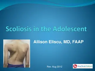
Understanding Scoliosis: Causes, Symptoms, and Diagnosis
Scoliosis is a condition where the spine curves laterally, often without causing pain. Severe cases may lead to nerve impingement or impact on vital organs. Understanding the biomechanics of scoliosis involves visualizing the spine in 3D planes. Clinical manifestations include rib prominence, leg length discrepancies, and potential pain and stiffness due to structural and functional impacts. The condition requires thorough differential diagnosis to rule out other potential causes such as tumors or spinal defects.

Download Presentation

Please find below an Image/Link to download the presentation.
The content on the website is provided AS IS for your information and personal use only. It may not be sold, licensed, or shared on other websites without obtaining consent from the author. Download presentation by click this link. If you encounter any issues during the download, it is possible that the publisher has removed the file from their server.
E N D
Presentation Transcript
Scoliosis By Prof. Salah Hawas
General description The word scoliosis is taken from the Greek language, translated to mean curvature. By definition, scoliosis refers to the spine curving laterally, either to the right or left side, and can be seen in childhood or adulthood. A normal spine viewed from behind the patient (posteroanterior [PA] view on radiograph) appears straight from the neck down to the buttocks. A scoliotic spine viewed from behind will resemble the shape of an S or C curve.
General description To study the biomechanics behind scoliosis, visualization of the spine in the three-dimensional (3-D) planes is required. To simplify the issue, one can imagine that as the spine curves to either the right or the left, the involved vertebrae must rotate to compensate for the curve to keep the body in balance.
General description The spine can curve abnormally mainly in two areas: the thoracic and/ or lumbar spine. If scoliosis occurs in the thoracic spine, the thoracic vertebrae, which are attached to the thoracic ribs, will rotate and result in rib prominence on the opposite side of the curve. If the scoliosis in the thoracic spine is very severe, it can lead to abnormal function of visceral organs within the thoracic cage, such as the heart and lungs.
Clinical manifestations Most authors believe that most patients with scoliosis never present with pain. It is only in severe cases (Cobb angles 70*), where nerve impingement or severe pulmonary (restriction in inhalation) and cardiac (corpulmonale) defects are involved, that scoliotic patients complain of pain.
Clinical manifestations As the spine curves laterally, the affected vertebrae rotate, affecting bone, ligament, and muscle connections to the spine. Rib prominence on the opposite side of the curve as well as leg length discrepancies are commonly seen with scoliosis. Because structure and function are highly integrated, these dysfunctional areas will ultimately lead to abnormal posture and strain on muscles, leading to pain and stiffness.
Differential diagnosis The differential diagnosis in a patient with scoliosis includes tumors, embryological defects (failure of segmentation of vertebrae or ribs), trauma, spinal stenosis, ankylosing spondylitis, and spondylolysis.
Etiologies The etiologies of scoliosis can be divided into three major categories: idiopathic, congenital vertebral, and neuromuscular defect. Each of these categories has many other subgroups, but idiopathic and congenital vertebral defects comprise the majority of presentations.
Infantile idiopathic scoliosis (03 years of age) Most patients with infantile idiopathic scoliosis present before 6 months of age with a curved spine that usually develops within the upper lumbar/ lower thoracic region. Most of these infants deformities revert to a normal curvature within the next few years.
Infantile idiopathic scoliosis (03 years of age) Diagnosis is usually made based on the upright PA and lateral views on radiograph. The treatment differs based on the degree of curvature. The Cobb angle is used to measure the amount of scoliosis. A person who has a Cobb angle 10 as determined by the X-ray (not with a scoliometer) is diagnosed as having scoliosis. The Cobb angle is determined by drawing a perpendicular line (on the X-ray) from the top most deviated vertebrae and the bottom most deviated vertebra. The two perpendicular lines meet to form the Cobb angle.
Treatment On the fi rst visit, treatment is usually not initiated. Most patients with an initial curve angle greater than 20 and an increase of 5 on the following offi ce visit should be treated aggressively (offi ce visits should be scheduled within 4 to 6 weeks). Treatment options include bracing or casting. If signs of scoliosis persist, an orthotic device must be worn 23 hours a day. Fusing the vertebra is usually not an option at this stage, although some physicians will insert a supporting rod if there is rapid progression.
Juvenile idiopathic scoliosis (310 years of age) History This group of patients manifests with signs beginning after age 3 but before age 10. Patients in this group are usually asymptomatic. Females make up most of these patients. Some authors believe that pain can be a symptom early on due to defects in walking habits. Unlike the infantile group, there seems to be a rapid progression of the deformity if not corrected early.
Juvenile idiopathic scoliosis (310 years of age) Physical During the examination, the physician should assess the symmetry among different body limbs, including shoulders, leg lengths, anterior superior iliac spine, posterior superior iliac spine, ischial tuberosities, and pubic symphysis. Skin texture and discoloration should be examined as well to rule out any neuromuscular defects. Neurological exams should be conducted to assess for any deficits.
Juvenile idiopathic scoliosis (310 years of age) Physical The Adams test assesses the paravertebral region of the patient. The patient stands with feet together and is asked to fl ex forward until at a 90* angle. Extreme paravertebral rotation and other irritant anomalies can become apparent during examination. The use of a scoliometer is not recommended for diagnosis due to the lack of experience of most physicians with the device.
Juvenile idiopathic scoliosis (310 years of age) Diagnosis Diagnosis should be made with radiographs of the lateral and PA views. Patients with a Cobb angle 10* based on the fi lm (not with a scoliometer) are diagnosed with scoliosis. Patients in this age group should also have radiographs of the brain to exclude other spinal pathologies.
Juvenile idiopathic scoliosis (310 years of age) Treatment Initial curvatures of Cobb angle 20* should be treated aggressively if the patient presents on the following visit with an increase of 5* in curvature within the next 4 6 months (increase in curvature is about 1* a month). If the patient presents with a curvature >25* on the fi rst visit, the physician might want to reexamine the patient earlier than the normal 4-month follow-up visit to prevent further increase in curvature.
Juvenile idiopathic scoliosis (310 years of age) Treatment Treatment in this patient population involves bracing the individual. If the patient progresses rapidly, surgery can be considered (although not done in most cases). Surgery consists of fusing the vertebra together. Most physicians don t consider this a viable option, as growth of the spine is prevented.
Adolescent idiopathic scoliosis (10 years of age to maturity) This age cohort accounts for most of the scoliosis seen by physicians (about 80%). The onset is usually manifested by puberty but before maturity. The majority of patients with adolescent onset are girls (92%).
Adolescent idiopathic scoliosis (10 years of age to maturity) History Most of these individuals do not have any symptoms and their deformities are recognized by an examiner, be it a parent, a primary care physician, or a school nurse. Most of these patients have a convex curve to the right in the thoracic region. Neurological deficits should be considered, especially in left-sided convex curves. Further imaging studies should be performed on these patients to check for intraspinal pathologies (neurofibroma, astrocytoma).
Adolescent idiopathic scoliosis (10 years of age to maturity) Physical and diagnosis The physician should conduct all of the tests used to assess juvenile idiopathic scoliosis for examination and diagnosis.
Adolescent idiopathic scoliosis (10 years of age to maturity) Treatment Treatment for this cohort has to be based on the level of skeletal maturity and the expected progression of the curve. Most curves won t have an angular increase if they have already hit menarche. In some instances, however, there will be a progression mostly with curves >40* even after menarche. Females have a much greater tendency to have curve progression than males (10:1).
Adolescent idiopathic scoliosis (10 years of age to maturity) Treatment Most patients with adolescent idiopathic scoliosis do not require treatment. Individuals with Cobb angle 20* with a progression of 5* should be treated. Most physicians use the Milwaukee brace as the primary treatment. The brace has to be worn for most of the day (23 hours; it may be removed for bathing only). Electrical stimulation has been used as an adjuvant. Surgery is only indicated once bracing has failed and the curve has progressed to 50*.


")
")