Mycobacterium Tuberculosis

 undefined


ALI M SOMILY MD
KING SAUD UNIVERSITY, COLLEGE OF
MEDICINE
Mycobacterium Tuberculosis
Objectives
Recognize that tuberculosis as
a chronic
disease
mainly affecting the respiratory system.
Know the
epidemiology
of tuberculosis world wide
and in the kingdom of Saudi Arabia
Understand the methods of
transmission
of
tuberculosis and the people at risk.
Know the causative agents and their
characteristic
and classification and
methods
of detection.
Understand the
pathogenesis
of tuberculosis.
Objectives
Differentiate between
primary and secondary
tuberculosis and the clinical features of each.
Understand the method of
tuberculin skin
test and
result interpretation.
Know the
laboratory and radiological
diagnostic
methods.
Know the
chemotherapeutic
and other methods of
management of tuberculosis cases.
Describe the methods of
prevention and control
of
tuberculosis.
Introduction
Tuberculosis (TB) is an ancient, chronic disease
affects humans, caused by Mycobacterium
tuberculosis complex.
A major cause of death worldwide.
Usually affects the lungs, other organs can be
affected in one third of cases.
If properly treated is curable, but fatal if untreated in
most cases.
Epidemiology
TB affects 1/3 of human race ( 2 billions) as a latent
dormant tuberculosis.
Incidence: a world wide disease, more common in
developing countries.
Affects all age groups who are subject to get the infection.
The WHO estimated 8.9 million new cases in 2004 &
million death.
Incidence :
KSA : 32-64 cases /100,000
USA : 5.2 cases/100,000
South Ease Africa : 290 cases /10,000 due to coupling with HIV
infection.
Estimated TB Incident Rate in 2014
Epidemiology
Transmission mainly through inhalation of airborne
droplet nuclei ( < 5 μm) in pulmonary diseases case,
rarely through GIT & skin
Reservoir: patients with open TB.
Age: young children & adults
People at risk : lab. technicians, workers in mines,
doctors, nurses.
HIV pts., diabetics end stage renal failure, contacts
with index case.
Characteristics of the Genus Mycobacteria
Slim, rod shaped, non-motile, do not form spores.
Do not stain by Gram stain. Why ?
Contain high lipid conc. ( Mycolic acid ) in the cell
wall which resist staining.
It is called Acid- alcohol fast bacili (AFB), Why ? It
resists de-colorization with up to 3% HCL,
5%ethanol or both.
Mycobacterium Cell Wall Structure
Acid Fast Stain
Acid- alcohol fast bacili
Acid-Fast Bacilli (AFB)
Stain used : Ziehl-Neelsen stain (ZN stain)
Strict aerobe
Multiply intracellularily
Delayed hypersensitivity reaction type of immune
response
Slowly growing ( wks.)
Mycobacterium tuberculosis complex
1-
M.tuberculosis
(Human type)
2-
M. bovis
(Bovine type)
3-
M. Africanum
4- BCG strains All are called Mycobacterium
tuberculosis Complex and cause tuberculosis ( TB).
Pathogenesis of Tuberculosis
Mycobacteria acquired by airborne droplet reaches
the alveolar macrophages, able to survive their (
main virulence factor).
This starts cell mediated immune response which
controls the multiplication of the organism but does
not kill it.
Granuloma formed, organism lives in dormant state
( latent tuberculosis infection)
Pathogenesis of Tuberculosis
Patient show evidence of delayed cell
mediated immunity ( CMI ).
Disease results due to destructive effect of
CMI.
Clinically the disease is divided into primary
or secondary.
Primary Tuberculosis occurs in patients not
previously infected.
Inhalation of bacilli
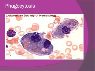
Phagocytosis lymph nodes calcify to produce
GHON focus (or Primary Complex) at the
periphery of mid zone of lung.
Stages of Tuberculosis
Histopathology
Micrographs of tissue specimens from patients with tuberculosis Low-power micrograph (×100) of a haematoxylin and eosin stained tissue section
from an immunocompetent patient with tuberculosis that shows a well formed tuberculous granuloma with a central area of caseous necrosis surrounded by
epithelioid macrophages, giant cells, and T lymphocytes, and surrounding outer fibrosis (A). Medium-power (×250) micrograph of tissue from a patient
infected with HIV with advanced immunodeficiency that shows mononuclear infiltrate but absence of granuloma formation (B). High-power (×400) tissue
section from the same specimen from the patient with advanced immunodeficiency with Ziehl-Neelsen staining showing numerous acid-fast bacilli with
little evidence of a cellular immune host response (C). Micrographs provided by Colleen Wright, Stellenbosch University, South Africa.
Radiology
Primary Tuberculosis
Genitourinary TB
Miliary TB (blood and other organs)
Soft tissue (cold abscess): lack of inflammation with
caseation.
Caseation: due to delayed hypersensitivity reaction.
Contains many bacilli, enzymes, O 2,N 2
intermediates, necrotic center of granuloma cheezy
material.
Secondary TB (reactivation)
Occurs later in life
Lung more common site
Immunocompromised patients.
Lesion localized in apices
Infectious & symptomatic
Microscopy: many bacilli, large area of caseous
necrosis cavity (open TB) with granuloma and
caseation.
Secondary TB
Clinically: fever, cough, hemoptysis, weight loss &
weakness.
Source of secondary TB : -
Endogenous (reactivation of an old TB)
or
Exogenous (re-infection in a previously sensitized patient who
has previous infection with the organism).
Immunity to Tuberculosis
Cell-mediated immunity associated with delayed
hypersensitivity reaction.
Detected by tuberculin skin test.
Tuberculin test takes 2-10 weeks to react to
tuberculin and becomes positive.
Tuberculin Skin Test
Uses purified protein derivative (PPD).
Activity expressed by Tuberculin unit.
Activates synthesized lymphocytes to produce CMI
which appear as skin induration.
May not distinguish between active and past
infection except in an individual with recent contact
with infected case.
Low level activity induced by environmental
mycobacteria, previous vaccination.
Methods of Tuberculin Skin Test
Intradermal inoculation of 0.1 ml of PPD, 5TU.
Read after 48-72hrs.
Methods of tuberculin skin test :
1- Mantoux test.
2- Heaf test (screening).
Positive Tuberculin Skin Test
1- >5mm induration positive in :
Recent contact with active TB.
HIV or high risk for HIV
Chest X-ray consistent with healed TB.
2- > 10mm induration positive in:
IV drugs user
HIV seronegative patient.
Medical conditions eg. diabetes, malignancy.
Residents & employee at high risk
Patients from country with high incidence.
Children < 4yrs or exposed to adult high risk group.
Mycobacteriology lab. personnel.
Positive Tuberculin Test
3- >15 mm induration positive in : any persons
including those with no risk factors for TB.
Negative Tuberculin Skin Test
No induration, either due to:
No previous infection
Pre-hypersensitivity stage
Lost TB sensitivity with loss of antigen.
AIDS patients are anergic and susceptible to
infection.
Laboratory Diagnosis of TB
1- Specimens:
Pulmonary TB: 3 early morning sputum samples ( or
induced cough),or bronchial lavage, or gastric
washing (infants),…etc.
Cerebrospinal fluid ( CSF) ( TB meningitis)
3 early morning urine
Bone, joint aspirate
Lymph nodes, pus or tissues NOT swab.
Repeat sample.
Laboratory Diagnosis of TB
2- Direct microscopy of specimen :
Z-N or (Auramine ) stain.
3- Culture: the gold standard test for identification
and sensitivity.
Media used: Lowenstein-Jensen media (L J).
Media contains: eggs, asparagin, glycerol, pyruvate/
malachite green.
Laboratory Diagnosis of TB
Colonies appear in LJ media after 2-8 weeks as eugenic,
raised, buff, adherent growth enhanced by glycerol (MTB) or
by pyruvate (M.bovis).
Other media plus LJ media may be used:
Fluid media (middle Brook)
MGIT ( mycobacteria growth indicator test )
Automated methods :- eg. Bactec MGIT.
Measurement of interferon –gamma ( IF-
γ)
secreted from
sensitized lymphocytes challenged by the same mycobacterial
proteins in a patient previously exposed to disease, will
produce interferon gamma. Has a specific significance than
tuberculin skin test.
PCR: molecular test directly from specimen (CSF).
Identification
Morphology, growth at 37C % CO 2
Biochemical tests : Niacin production & Nitrate
test.
Sensitivity testing
Guinea pig inoculation: rarely done.
Management of a TB case
1- Isolation for days ( for smear positive cases i.e. >
1000 organisms / ml of sputum considered
infectious case ).
Triple regimen of therapy.Why ?
To prevent resistant mutants
To cover strains located at different sites of the lung.
To prevent relapse
2- Treatment must be guided by sensitivity testing.
First Line Treatment
Isoniazide (INH)
Rifampicin (RIF)
Ethmbutol (E)
Pyrazinamide (P)
Streptomycin (S) INH+ RIF +P for 2 months then
continue with INH+RIF for 4-6 months.
Multidrug resistant TB is resistance to INH & RIF.
Directly Observed Therapy (DOT).
Second Line
Second Line Used if the bacteria was resistant to first
line drugs.
More toxic than the first line drugs.
PASA ( Para-Amino Salicylic acid)
Ethionamide
Cycloserine,
Kanamycin,
Fluroquiolones
Prevention of TB
Tuberculin testing of herds.
Slaughter of infected animals.
Pasteurization of milk to prevent bovine TB
Recognition of new cases.
Prophylaxis with INH of contacts.
Follow up cases.
Immunization with BCG to all new borne.
Tuberculosis is a chronic disease caused by Mycobacterium tuberculosis that primarily affects the respiratory system. This overview covers the epidemiology, transmission methods, causative agents, pathogenesis, diagnostic methods, management, prevention, and control of tuberculosis. The importance of recognizing primary and secondary tuberculosis, along with clinical features and interpretation of diagnostic tests, is also discussed. With a focus on raising awareness about this global health issue, the content aims to provide a comprehensive understanding of tuberculosis for healthcare professionals and the general public.

Download Presentation

Please find below an Image/Link to download the presentation.
The content on the website is provided AS IS for your information and personal use only. It may not be sold, licensed, or shared on other websites without obtaining consent from the author. Download presentation by click this link. If you encounter any issues during the download, it is possible that the publisher has removed the file from their server.
E N D
Presentation Transcript
Mycobacterium Tuberculosis ALI M SOMILY MD KING SAUD UNIVERSITY, COLLEGE OF MEDICINE
Objectives Recognize that tuberculosis as a chronic disease mainly affecting the respiratory system. Know the epidemiology of tuberculosis world wide and in the kingdom of Saudi Arabia Understand the methods of transmission of tuberculosis and the people at risk. Know the causative agents and their characteristic and classification and methods of detection. Understand the pathogenesis of tuberculosis.
Objectives Differentiate between primary and secondary tuberculosis and the clinical features of each. Understand the method of tuberculin skin test and result interpretation. Know the laboratory and radiological diagnostic methods. Know the chemotherapeutic and other methods of management of tuberculosis cases. Describe the methods of prevention and control of tuberculosis.
Introduction Tuberculosis (TB) is an ancient, chronic disease affects humans, caused by Mycobacterium tuberculosis complex. A major cause of death worldwide. Usually affects the lungs, other organs can be affected in one third of cases. If properly treated is curable, but fatal if untreated in most cases.
Epidemiology TB affects 1/3 of human race ( 2 billions) as a latent dormant tuberculosis. Incidence: a world wide disease, more common in developing countries. Affects all age groups who are subject to get the infection. The WHO estimated 8.9 million new cases in 2004 & million death. Incidence : KSA : 32-64 cases /100,000 USA : 5.2 cases/100,000 South Ease Africa : 290 cases /10,000 due to coupling with HIV infection.
Epidemiology Transmission mainly through inhalation of airborne droplet nuclei ( < 5 m) in pulmonary diseases case, rarely through GIT & skin Reservoir: patients with open TB. Age: young children & adults People at risk : lab. technicians, workers in mines, doctors, nurses. HIV pts., diabetics end stage renal failure, contacts with index case.
Characteristics of the Genus Mycobacteria Slim, rod shaped, non-motile, do not form spores. Do not stain by Gram stain. Why ? Contain high lipid conc. ( Mycolic acid ) in the cell wall which resist staining. It is called Acid- alcohol fast bacili (AFB), Why ? It resists de-colorization with up to 3% HCL, 5%ethanol or both.
Acid-Fast Bacilli (AFB) Stain used : Ziehl-Neelsen stain (ZN stain) Strict aerobe Multiply intracellularily Delayed hypersensitivity reaction type of immune response Slowly growing ( wks.)
Mycobacterium tuberculosis complex 1- M.tuberculosis (Human type) 2- M. bovis (Bovine type) 3- M. Africanum 4- BCG strains All are called Mycobacterium tuberculosis Complex and cause tuberculosis ( TB).
Pathogenesis of Tuberculosis Mycobacteria acquired by airborne droplet reaches the alveolar macrophages, able to survive their ( main virulence factor). This starts cell mediated immune response which controls the multiplication of the organism but does not kill it. Granuloma formed, organism lives in dormant state ( latent tuberculosis infection)
Pathogenesis of Tuberculosis Patient show evidence of delayed cell mediated immunity ( CMI ). Disease results due to destructive effect of CMI. Clinically the disease is divided into primary or secondary. Primary Tuberculosis occurs in patients not previously infected. Inhalation of bacilli Phagocytosis lymph nodes calcify to produce GHON focus (or Primary Complex) at the periphery of mid zone of lung.
Histopathology Micrographs of tissue specimens from patients with tuberculosis Low-power micrograph ( 100) of a haematoxylin and eosin stained tissue section from an immunocompetent patient with tuberculosis that shows a well formed tuberculous granuloma with a central area of caseous necrosis surrounded by epithelioid macrophages, giant cells, and T lymphocytes, and surrounding outer fibrosis (A). Medium-power ( 250) micrograph of tissue from a patient infected with HIV with advanced immunodeficiency that shows mononuclear infiltrate but absence of granuloma formation (B). High-power ( 400) tissue section from the same specimen from the patient with advanced immunodeficiency with Ziehl-Neelsen staining showing numerous acid-fast bacilli with little evidence of a cellular immune host response (C). Micrographs provided by Colleen Wright, Stellenbosch University, South Africa.
Primary Tuberculosis Genitourinary TB Miliary TB (blood and other organs) Soft tissue (cold abscess): lack of inflammation with caseation. Caseation: due to delayed hypersensitivity reaction. Contains many bacilli, enzymes, O 2,N 2 intermediates, necrotic center of granuloma cheezy material.
Secondary TB (reactivation) Occurs later in life Lung more common site Immunocompromised patients. Lesion localized in apices Infectious & symptomatic Microscopy: many bacilli, large area of caseous necrosis cavity (open TB) with granuloma and caseation.
Secondary TB Clinically: fever, cough, hemoptysis, weight loss & weakness. Source of secondary TB : - Endogenous (reactivation of an old TB) or Exogenous (re-infection in a previously sensitized patient who has previous infection with the organism).
Immunity to Tuberculosis Cell-mediated immunity associated with delayed hypersensitivity reaction. Detected by tuberculin skin test. Tuberculin test takes 2-10 weeks to react to tuberculin and becomes positive.
Tuberculin Skin Test Uses purified protein derivative (PPD). Activity expressed by Tuberculin unit. Activates synthesized lymphocytes to produce CMI which appear as skin induration. May not distinguish between active and past infection except in an individual with recent contact with infected case. Low level activity induced by environmental mycobacteria, previous vaccination.
Methods of Tuberculin Skin Test Intradermal inoculation of 0.1 ml of PPD, 5TU. Read after 48-72hrs. Methods of tuberculin skin test : 1- Mantoux test. 2- Heaf test (screening).
Positive Tuberculin Skin Test 1- >5mm induration positive in : Recent contact with active TB. HIV or high risk for HIV Chest X-ray consistent with healed TB. 2- > 10mm induration positive in: IV drugs user HIV seronegative patient. Medical conditions eg. diabetes, malignancy. Residents & employee at high risk Patients from country with high incidence. Children < 4yrs or exposed to adult high risk group. Mycobacteriology lab. personnel.
Positive Tuberculin Test 3- >15 mm induration positive in : including those with no risk factors for TB. any persons
Negative Tuberculin Skin Test No induration, either due to: No previous infection Pre-hypersensitivity stage Lost TB sensitivity with loss of antigen. AIDS patients are anergic and susceptible to infection.
Laboratory Diagnosis of TB 1- Specimens: Pulmonary TB: 3 early morning sputum samples ( or induced cough),or bronchial lavage, or gastric washing (infants), etc. Cerebrospinal fluid ( CSF) ( TB meningitis) 3 early morning urine Bone, joint aspirate Lymph nodes, pus or tissues NOT swab. Repeat sample.
Laboratory Diagnosis of TB 2- Direct microscopy of specimen : Z-N or (Auramine ) stain. 3- Culture: the gold standard test for identification and sensitivity. Media used: Lowenstein-Jensen media (L J). Media contains: eggs, asparagin, glycerol, pyruvate/ malachite green.
Laboratory Diagnosis of TB Colonies appear in LJ media after 2-8 weeks as eugenic, raised, buff, adherent growth enhanced by glycerol (MTB) or by pyruvate (M.bovis). Other media plus LJ media may be used: Fluid media (middle Brook) MGIT ( mycobacteria growth indicator test ) Automated methods :- eg. Bactec MGIT. Measurement of interferon gamma ( IF- ) secreted from sensitized lymphocytes challenged by the same mycobacterial proteins in a patient previously exposed to disease, will produce interferon gamma. Has a specific significance than tuberculin skin test. PCR: molecular test directly from specimen (CSF).
Identification Morphology, growth at 37C % CO 2 Biochemical tests : Niacin production & Nitrate test. Sensitivity testing Guinea pig inoculation: rarely done.
Management of a TB case 1- Isolation for days ( for smear positive cases i.e. > 1000 organisms / ml of sputum considered infectious case ). Triple regimen of therapy.Why ? To prevent resistant mutants To cover strains located at different sites of the lung. To prevent relapse 2- Treatment must be guided by sensitivity testing.
First Line Treatment Isoniazide (INH) Rifampicin (RIF) Ethmbutol (E) Pyrazinamide (P) Streptomycin (S) INH+ RIF +P for 2 months then continue with INH+RIF for 4-6 months. Multidrug resistant TB is resistance to INH & RIF. Directly Observed Therapy (DOT).
Second Line Second Line Used if the bacteria was resistant to first line drugs. More toxic than the first line drugs. PASA ( Para-Amino Salicylic acid) Ethionamide Cycloserine, Kanamycin, Fluroquiolones
Prevention of TB Tuberculin testing of herds. Slaughter of infected animals. Pasteurization of milk to prevent bovine TB Recognition of new cases. Prophylaxis with INH of contacts. Follow up cases. Immunization with BCG to all new borne.


")


")