Fundamentals of Microscopy in Microbiology: An Insightful Journey
Explore the intriguing world of microscopy in microbiology, delving into the history, magnification, resolution, numerical aperture, and focal length. Uncover the evolution of microscopes and the fundamental concepts that underpin the science of microscopy. Gain a deeper understanding of how these tools enable researchers to visualize and study the unseen microcosm.

Download Presentation

Please find below an Image/Link to download the presentation.
The content on the website is provided AS IS for your information and personal use only. It may not be sold, licensed, or shared on other websites without obtaining consent from the author. Download presentation by click this link. If you encounter any issues during the download, it is possible that the publisher has removed the file from their server.
E N D
Presentation Transcript
B. Sc. First Year Semester I Paper I Fundamentals of Microbiology Unit 4: MICROSCOPY Dr. Jadhav P. N. Professor and Head, Microbiology Department, Deogiri College, Aurangabad
Introduction A microscope (Greek: mikron = small and scopeos = to look). MICROSCOPE: Is an instrument for viewing objects that are too small to be seen by the naked or unaided eye. MICROSCOPY: The science of investigating small objects using such an instrument is called microscopy.
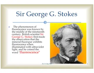
History 1590 - Hans Janssen and his son Zacharias Janssen, developed first microscope. 1609 - Galileo Galilei - occhiolino or compound microscope. 1620 - Christian Huygens, another Dutchman, developed a simple 2-lens ocular system that was chromatically corrected.
Magnification Degree of enlargement. No of times the length, breadth or diameter, of an object is multiplied. It depends upon Optical tube length Focal length of objective Magnifying power of eye piece TOTAL MAGNIFICATION: magnification of the eyepiece x magnification of the objective
Resolution Ability to reveal closely adjacent structural details as separate and distinct. LIMIT OF RESOLUTION (LR): The minimum distance between two visible bodies at which they can be seen as separate and not in contact with each other. LR = (0.61 x W)/NA W = Wavelength; NA = Numerical aperature
Numerical Aperture Ratio of diameter of lens to its focal length NA = n Sin /2 n = refractive index, = angle of aperture Definition: Capacity of an objective to render outline of the image of an object clear and distinct.
Focal length The term focal length refers to the amount of distance required between the objective lens and the top of your object, in order to be able to view an image through the microscope that is in-focus. When using a biological microscope the higher your objective magnification, the shorter the focal length generally is. With the 4x objective there may be some space between the objective lens and your cover slip on your prepared slide. However, when using the 100x objective lens the objective will be almost touching the cover slip, due to a smaller focal length.
Objective lens Low Power Objective (10x) This objective lens is the next lowest powered and is often the most helpful when it comes to analyzing glass slide samples. The total magnification for this lens is equal to 100x magnification (10x eyepiece lens x the 10x objective equals 100). Since it still provides a good amount of magnification at a good distance from the slide, there is a limited risk of it breaking the glass and potentially ruining the sample. Hence, why it is often preferred before going for a high powered lens. High Power Objective Lens (40x) This is referred to as the high powered objective lens since it is ideal for observing the small details within a specimen sample. The total magnification for this lens is equal to 400x magnification (10x eyepiece lens x the 40x objective equals 400). Oil Immersion Objective (100x) This objective lens will achieve the greatest magnification and has a total magnification of 1000x (10x eyepiece lens x the 100x objective equals 1000). However, since the refractive index of air and the glass slide are slightly different, a special oil must be used to help fill the gap between the two. Without a drop of oil, the objective lens will not work properly and you will not achieve the desired magnification and resolution.
Oculars At the top of the body tube, a lens is planted which is known as the eyepiece. On the rim of the eyepiece, there are certain markings such as 5X, 10X, 15X, etc. Which indicates the magnification power. The object s magnified image can be observed with the help of an eyepiece. The simplest negative eyepiece design, often termed the Huygenian eye-piece, is found on most teaching and laboratory microscopes fitted with achromatic objectives. Ramsden eyepiece has an eye lens and field lens that are also plano-convex, but the field lens is mounted with the curved surface facing towards the eye lens.
Condensers The condenser or sub-stage condenser is located between the light source and the stage. It has a series of lenses to converge on the object, light rays coming from the light source. After passing through the object, the light rays enter into the objective. The light condensing , light converging or light gathering capacity of a condenser is called numerical aperture of the condenser . Similarly, the light gathering capacity of an objective is called numerical aperture of the objective . If the condenser converges light in a wide angle, its numerical aperture is greater and vice versa. Most common condensers have numerical aperture 1.25.
There are three types of condensers as follows: (a) Abbe condenser (Numerical aperture=1.25): It is extensively used. (b) Variable focus condenser (Numerical aperture =1.25) (c) Achromatic condenser (Numerical aperture =1.40): It has been corrected for both spherical and chromatic aberration and is used in research microscopes and photomicrographs.
Iris diaphragm If light coming from the light source is brilliant and all the light is allowed to pass to the object through the condenser, the object gets brilliantly illuminated and cannot be visualized properly. Therefore, an iris diaphragm is fixed below the condenser to control the amount of light entering into the condenser.