Laboratory Diagnosis of Malaria: Methods and Procedures
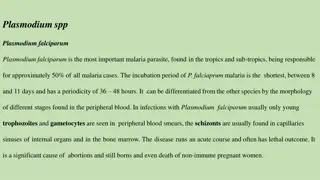
Methods for diagnosing malaria in laboratory settings involve techniques such as light microscopy and rapid diagnostic tests. Light microscopy is used to prepare blood films, enabling the detection of malaria parasites like Plasmodium falciparum. Rapid diagnostic tests can quickly identify malaria antigens. Understanding the morphology of malaria parasites, including trophozoites, schizonts, and gametocytes, is crucial for accurate diagnosis.

Download Presentation

Please find below an Image/Link to download the presentation.
The content on the website is provided AS IS for your information and personal use only. It may not be sold, licensed, or shared on other websites without obtaining consent from the author. Download presentation by click this link. If you encounter any issues during the download, it is possible that the publisher has removed the file from their server.
E N D
Presentation Transcript
PRACTICAL PRACTICAL ON BLOOD ON BLOOD PARASITES PARASITES
Common methods for parasitological diagnosis of malaria The two methods common in use : 1: Light microscopy 2: Rapid diagnostic tests (RDTs).
Laboratory diagnosis of malaria Laboratory diagnosis of malaria Rapid diagnostic tests detect Rapid diagnostic tests detect malaria antigens malaria antigens
Laboratory diagnosis of malaria Light microscopy:1: Preparing blood film
Light microscopy:1: Preparing blood film Laboratory diagnosis of malaria
Laboratory diagnosis of malaria Light microscopy: Thick and thin films
Laboratory diagnosis of malaria Plasmodium falciparum (trophozoite stage in thick smear) Plasmodium falciparum (trophozoite stage in thin smear) CCMOV BD
16 Morphology of Malaria The Malaria Parasite Trophozoites Three developmental stages seen in blood films: CCMOVBD CCMOVBD 1. Trophozoite Schizont Gametocyte 2. Schizont 3. Gametocyte CCMOVBD CCMOVBD
17 Morphology of Malaria Plasmodium vivax CCMOVBD Species of malaria is identified by its chracteristic microscopic appearance: Plasmodium ovale Plasmodium malariae Malaria Tutorials, Wellcome Trust CCMOVBD
18 Morphology of Malaria Plasmodium falciparum (trophozoite stage in thin smear) CCMOVBD
19 Morphology of Malaria Plasmodium falciparum (trophozoite stage in thick smear) CCMOVBD
20 Morphology of Malaria Plasmodium falciparum (characteristic banana-shaped or crescent shaped gametocyte stage in thin smear) CCMOVBD
A 25 year-old male from India, who came 3 months ago was admitted in KKUH with a history of severe anaemia and intermittent high grade fever for the last two months not responding to antibiotics. WHAT IS THE DIAGNOSIS? Diagnosis: malaria or Plasmodium vivax
A businessman who makes frequent trips to Thailand , presents with intermittent fever . WHAT IS THE DIAGNOSIS? Diagnosis: malaria or Plasmodium vivax
A student in KSU who returned three weeks from vacation in Africa , he developed intermittent fever last week and lost consciousness a short time ago. WHAT IS THE DIAGNOSIS? Diagnosis: malaria or Plasmodium falciparum
The patient was then treated with schizontocidal antimalarial drugs, a follow-up blood film is shown . ARE THERE ANY PARASITES? WHAT STAGE ? Plasmodium flaciparum , gametocyte stage
A 7 year old child presented with anemia , hepatospenomegaly and fever .Not responding to antimalarials and antibiotics . Bone marrow smear is shown ARE THERE ANY PARASITES? WHAT STAGE ? Leishmania , amastigote stage
Bone marrow aspiration Bone marrow amastigotes