Cell Communication and Signalling in Biology

Cells communicate through extracellular signalling molecules that bind to specific receptors on target cells, leading to changes in cellular behavior. Hormones such as steroid and peptide hormones, neurotransmitters like nor-adrenaline and acetylcholine, play crucial roles in cell signaling. Receptor proteins for these molecules can be located either inside the cell or on the cell membrane surface, depending on the hydrophobic or hydrophilic nature of the signal. Hydrophobic signals, like steroid hormones, control transcription by interacting with intracellular receptors to initiate gene expression.

Download Presentation

Please find below an Image/Link to download the presentation.
The content on the website is provided AS IS for your information and personal use only. It may not be sold, licensed, or shared on other websites without obtaining consent from the author. Download presentation by click this link. If you encounter any issues during the download, it is possible that the publisher has removed the file from their server.
E N D
Presentation Transcript
1.4 Communication and Signalling Advanced Higher Biology
Definitions for this Key Area What is an extracellular signalling molecule? What is a target cell? What is a receptor molecule? What happens when a signal molecule binds to a receptor molecule? What is a hormone? Name two types of hormone and give examples What is a neurotransmitter? Where are receptor proteins located? Why? How do hydrophobic signals control transcription?
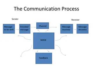
Coordination Cells must be able to communicate with each other. They do this through extracellular signalling molecules which are released from one cell and attach to a specific receptor on a target cell. The signalling molecule should change the conformation of the receptor, and thus, the behaviour of the target cell in some way. Different cell types produce specific signals that can only be detected and responded to by cells with the specific receptor
Examples of Extracellular Signalling Molecules Steroid hormones Peptide hormones Neurotransmitters
Hormones Secreted by tissue such as an endocrine gland Moves through bloodstream to target organ, where it joins with a receptor protein. Hormones can be either: Hydrophobic such as thyroxine or steroids Hydrophillic such as ADH or insulin
Neurotransmitters Extracellular signalling molecules Moves through the synapse from one neuron to another. Faster and more specific than hormones Includes nor-adrenaline and acetylcholine
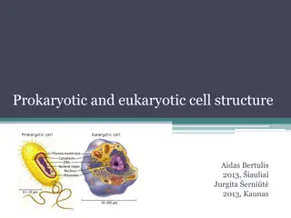
Location of Receptor Proteins Hydrophobic signalling molecules can pass through the cell membrane by diffusion as this is also hydrophobic. The receptors for this type of molecule are inside the cell in the cytoplasm or nucleus Hydrophilic signalling molecules can t get through the cell membrane so require a receptor on the cell membrane surface.
Hydrophobic Signals and the Control of Transcription Steroid hormones cause transcription to take place. When a steroid reaches the receptor inside the cell, it causes a conformational change in the receptor which turns ON transcription. In more detail: 1. Steroid hormone binds to specific receptor (in cytosol or nucleus) 2. This hormone receptor complex then moves to the nucleus and binds with specific DNA sequences called Hormone Response Elements 3. This influences the rate of transcription, with each steroid affecting the expression of many genes
Signal Transduction Hydrophillic signalling molecules cannot pass through the cell membrane as they are not lipid soluble. Examples of hydrophillic signalling molecules are: Peptide hormones Neurotransmitters
Signal Transduction Since hydrophillic signalling molecules cannot pass through the cell membrane, they must join to a receptor on the outside of the cell membrane. When a signalling molecule binds to the receptor on the cell membrane, a conformational change happens. The signal molecule does not go into the cell, but the signal is transduced this means it is passed across the membrane and a response happens.
Signal Transduction Signal transduction often involves: Cascades activated by G-proteins Phosphorylation by kinase enzymes Examples of peptide hormones are: Insulin Glucagon Anti-diuretic hormone (ADH) Each peptide hormone has a specific receptor on the target cell surface, making them highly specific.
What is a G-Protein? Molecules which relay signals from activated receptors. An activated receptor is one which is bound to a signalling molecule
Phosphorylation A phosphorylation cascade allows more than one intracellular signalling pathway to be activated. What are they? A series of events where one kinase activates the next in the sequence and so on. Cascades result in the phosphorylation of many proteins as a result of one signalling event.
Insulin and the Recruitment of GLUT4 Glucose levels within the blood must be kept within 3.9 6.1mmol per litre. The peptide hormones insulin and glucagon control blood glucose level. If the blood glucose level rises, usually after a meal, the pancreas detects an increase in blood glucose. This causes insulin to be released
Insulin and the Recruitment of GLUT4 The insulin receptor is a kinase-linked receptor on the membrane of fat and muscle cells. If a hydrophillic signal binds to the insulin receptor, a signal is transduced and a series of phosphorylation events trigger the recruitment of GLUT4 glucose transporters to the cell membrane. Glucose is allowed to enter the cell, lowering it s concentration in the blood. The glucose in the cell is metabolised.
Diabetes Diabetes mellitus is caused by a deficiency in the effect of insulin and results in the loss of control of blood glucose level. Two types: Failure to produce insulin (type 1) Loss of receptor function (type 2)
Diabetes Type 1 diabetes is treated with injections of insulin it cannot be reversed. It is often genetic. Type 2 diabetes is reversible, as cells are no longer sensitive to insulin, normally due to obesity. Exercise and losing weight can reduce the impact of type 2 diabetes as it triggers recruitment of GLUT4 through other metabolic pathways. Exercise improves the uptake of glucose to fat and muscle cells.
Nerve Transmission As well as the sodium potassium pump, potassium ions also move in another way in some cells. In nerve cells (neurons), the cell membrane has a potassium channel which lets some of the potassium ions leak back out of the cell. This results in a net positive charge outside of the cell, and a net negative charge inside the cell.
Nerve Transmission This imbalance in the electrical charge across the neuron membrane is called the resting potential. A nerve impulse in the body is only passed along when this resting potential is depolarised (changed from negative inside the cell back to neutral/positive)
How are nerves triggered? Depolarisation of the resting potential is triggered by neurotransmitters at a synapse. The neurotransmitter (e.g. Acetylcholine) diffuses across the synapse and binds to a transmembrane receptor protein on the surface of the next neuron.
How are nerves triggered? This receptor is a ligand gated Sodium ion channel so the binding of the neurotransmitter causes a change in conformation on the neuron cell membrane, making the channel open and allowing the sodium ions to flood through.
How are nerves triggered? If lots of ions move through, then the voltage across the membrane reaches a level where it has been depolarised (moved from negative charge to neutral/positive) The change in voltage means each neighbouring voltage gated sodium channel opens up, leading to a domino effect (wave of depolarisation) through the nervous system. This is why the nervous system is a fast, electrical system.
How are nerves reset? There has to be a process to reset the resting potential, or nerves would be constantly stimulated and signals would be constantly sent through the body. This happens when the voltage inside of the cell reaches a critically high level.
How are nerves reset? The voltage gated sodium channels close and voltage gated potassium channels open. This is called re-polarisation.